Korean J Gastroenterol.
2017 Dec;70(6):304-307. 10.4166/kjg.2017.70.6.304.
Diagnosis and Removal of Ascaris lumbricoides during Endoscopic Examination
- Affiliations
-
- 1Department of Internal Medicine, Seoul National University College of Medicine, Seoul, Korea. kjwjor@snu.ac.kr
- 2Department of Internal Medicine, Seoul Metropolitan Government-Seoul National University Boramae Medical Center, Seoul, Korea.
- KMID: 2398882
- DOI: http://doi.org/10.4166/kjg.2017.70.6.304
Abstract
- No abstract available.
MeSH Terms
Figure
-

Fig. 1. Colonoscopic view of Ascaris lumbricoides that was found in the descending colon.
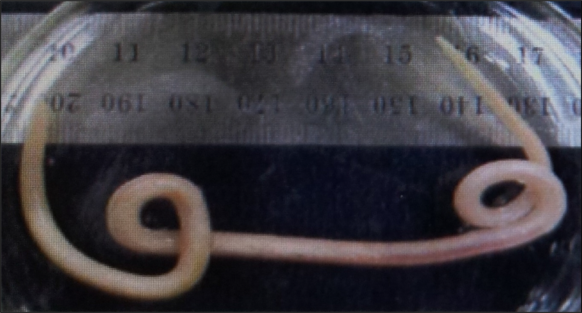
Fig. 2. Ascaris lumbricoides that was extracted from the colon.

Fig. 3. Three lips of the Ascaris lumbricoides that was extracted from the colon.
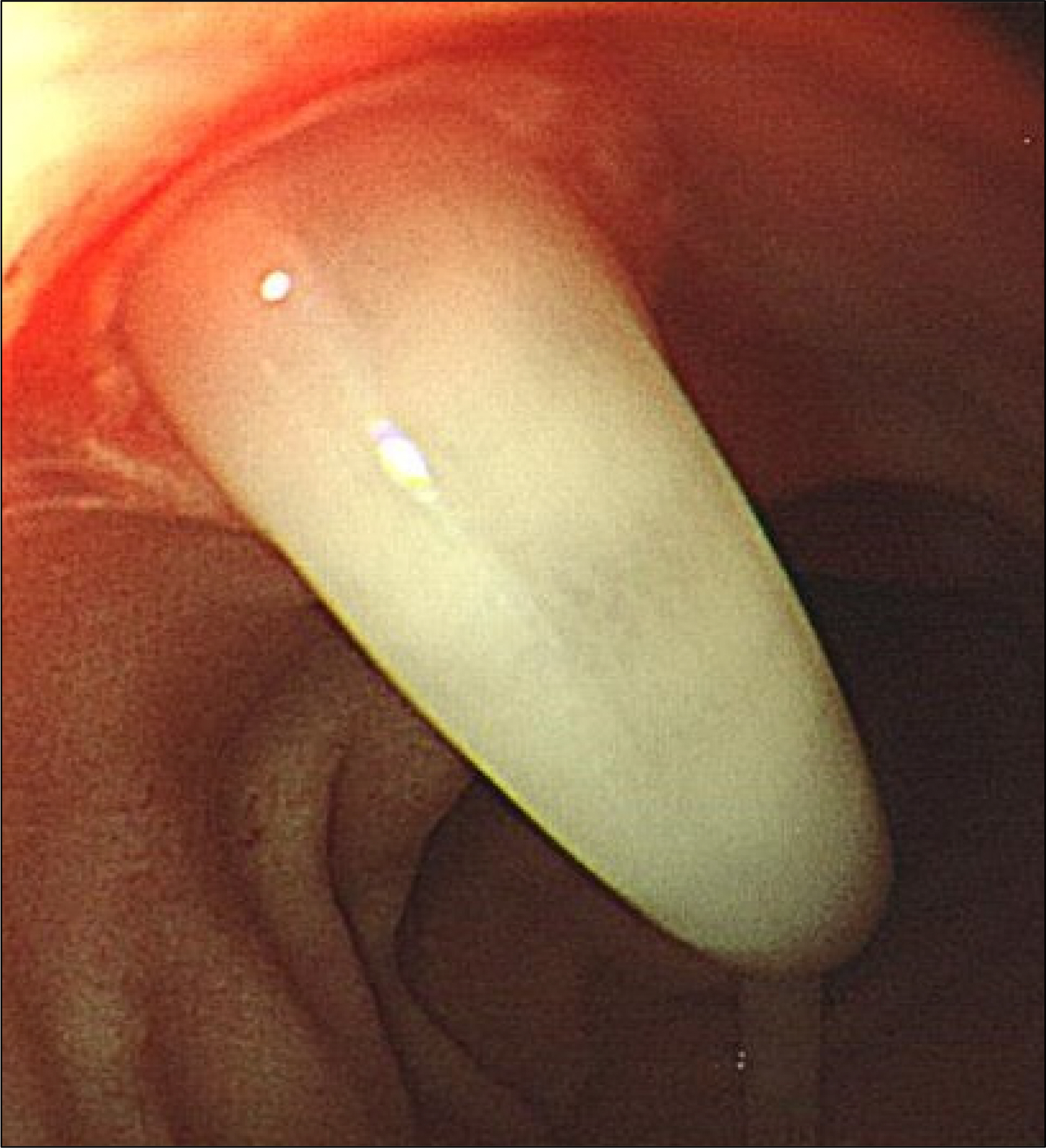
Fig. 4. Ascaris lumbricoides sticking out of the ampulla of Vater.
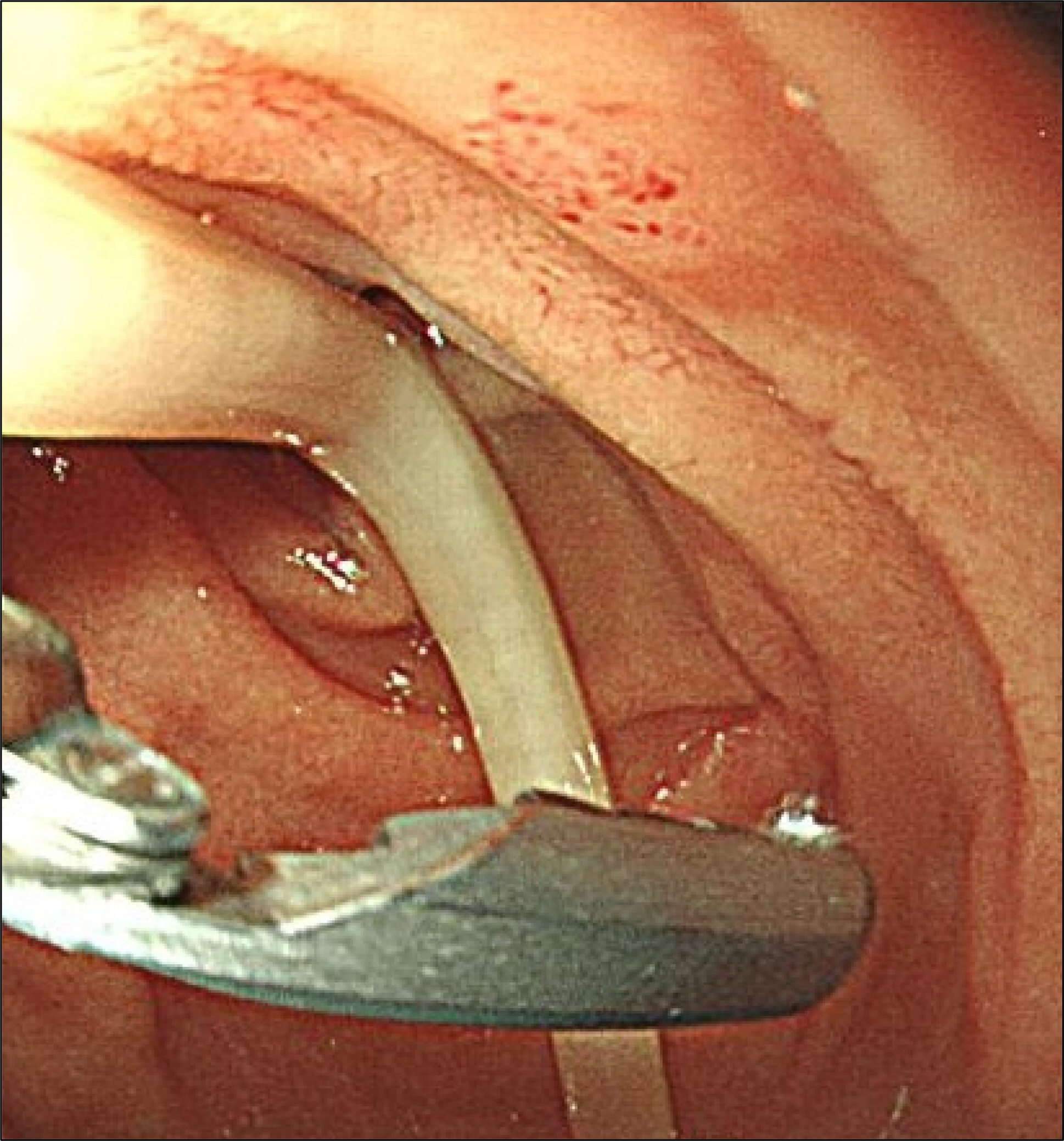
Fig. 5. Removal of Ascaris lumbricoides using alligator forceps.
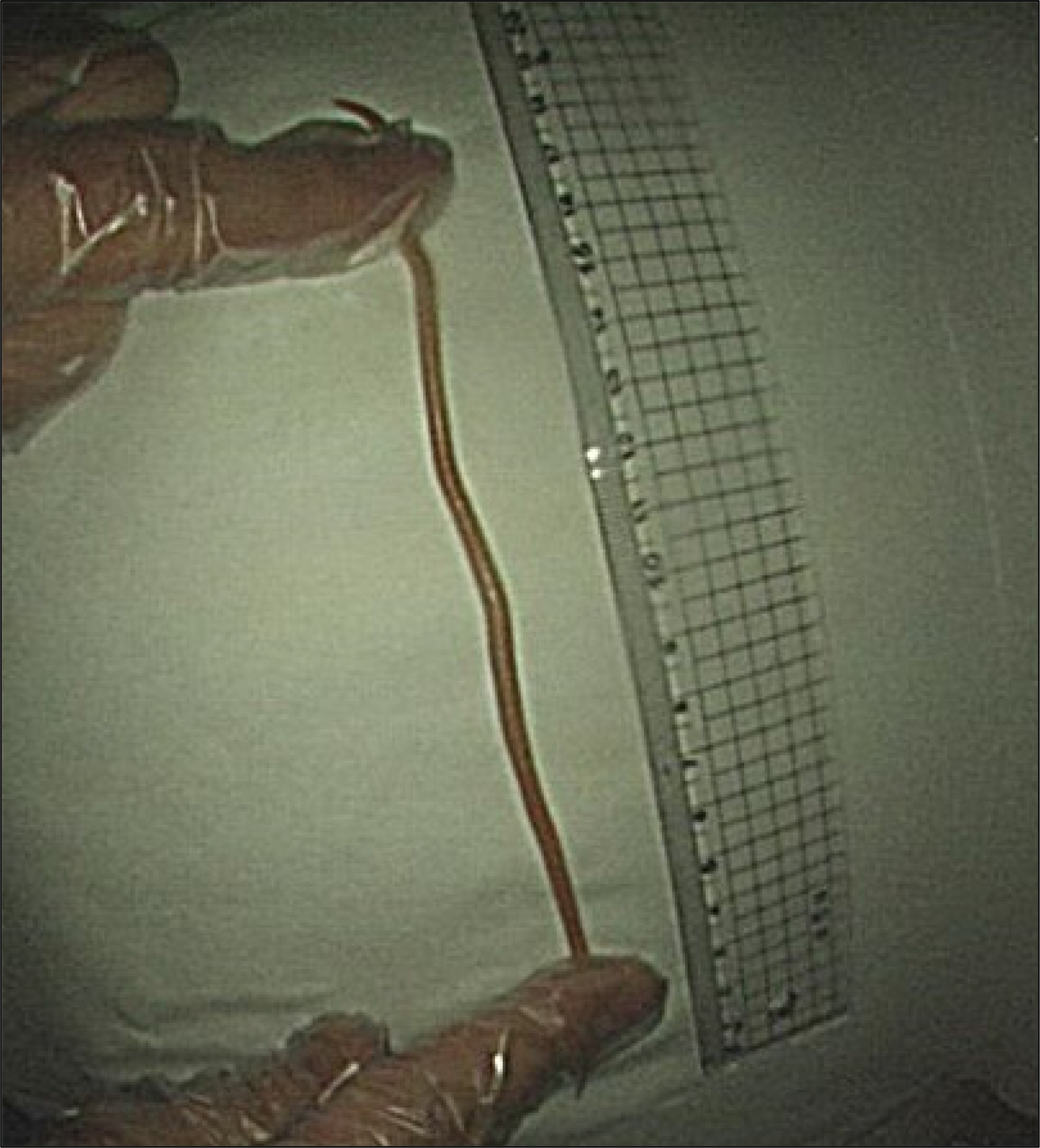
Fig. 6. The Ascaris lumbricoides measured 20 cm in length.
Reference
-
References
1. de Silva NR, Brooker S, Hotez PJ, Montresor A, Engels D, Savioli L. Soil-transmitted helminth infections: updating the global picture. Trends Parasitol. 2003; 19:547–551.
Article2. Kim TS, Cho SH, Huh S, et al. A nationwide survey on the abdominal of intestinal parasitic infections in the Republic of Korea, 2004. Korean J Parasitol. 2009; 47:37–47.3. National surveys of intestinal parasitic infections in Korea, 8th abdominal (2013). [Internet]. Cheongju: Korea Centers for Disease Control and Prevention;2014. Jan 29 [cited 2017 Nov 2]. Available from:. http://cdc.go.kr/CDC/info/CdcKrInfo0301.jsp?menuIds=HOME001-MNU1154-MNU0005-MNU0037&cid=24152.
- Full Text Links
-
- Actions
-
Cited
- CITED
-
- Close
- Share
-
- Similar articles
-
- A Case of Acute Pancreatitis due to Impaction of Ascaris lumbricoides into the Pancreatic Duct
- Biliary Ascariasis with Choledocholithiasis and Biliary Pancreatitis: Endoscopically Treated
- A Case of Ascarid Chronic Pancreatitis Due to Impaction of Ascaris Lumbricoides into the pancreatic Duct
- Studies on the intradermal reactions with the fractions of Ascaris lumbricoides
- Two Worms of Ascaris lumbricoides in the Bile Duct Combined with Bile duct Stones and Liver Abscess: A Case Report
