Comparative kinematic gait analysis in young and old Beagle dogs
- Affiliations
-
- 1Small Animal Clinic, Epidemiology and Information Processing, University of Veterinary Medicine Hannover, D-30559 Hannover, Germany. Ingo.Nolte@tiho-hannover.de
- 2Institute for Biometry, Epidemiology and Information Processing, University of Veterinary Medicine Hannover, D-30559 Hannover, Germany.
- 3Division of Medicine Clinic III, Hematology, Oncology and Palliative Medicine, University of Rostock, D-18057 Rostock, Germany.
- KMID: 2398512
- DOI: http://doi.org/10.4142/jvs.2017.18.4.521
Abstract
- Age-related involution in dogs involves loss of muscle mass and changes in connective tissue and articular cartilage. The aim of this study was to examine whether an age-related influence on joint mobility can be detected in the absence of disease. Five young (mean age 2.0 years) and five old (mean age 10.4 years) healthy and sound Beagle dogs underwent computer-assisted gait analysis during locomotion on a treadmill. Shoulder, elbow, carpal, hip, stifle, and tarsal joint angles including joint angle progression curves, minimum and maximum joint angles, and range of motion (ROM) in degrees were analyzed. The old group had a smaller maximum joint angle (p = 0.037) and ROM (p = 0.037) of the carpal joint; there were similar tendencies in the shoulder, elbow, and carpal joints. Descriptive analysis of the progression curves revealed less flexion and extension of the forelimb joints. The results indicate restricted joint mobility of the forelimb in old dogs, primarily of the carpal joint. Results in the joints of the hindlimb were inconsistent, and the contrasting alterations may be due to a compensatory mechanism. As most alterations were found in the distal joints, these should receive particular attention when examining elderly dogs.
Keyword
MeSH Terms
Figure
-
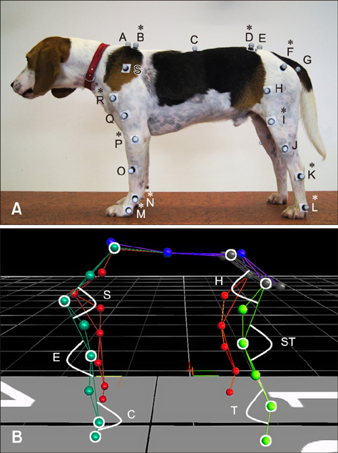
Fig. 1 Positions of the retroreflective passive markers on a representative Beagle dog (A) and the processed kinematic rod model (B). (A) Positions of joint-determining markers (asterisks) and additional markers. A, cervicothoracic transition; B, margo dorsalis scapulae; C, thoracolumbar transition; D, crista iliaca ossis illi; E, os sacrum; F, trochanter major ossis femoris; G, tuber ischiadicum ossis illi; H, femur; I, epicondylus lateralis ossis femoris; J, tibia; K, malleolus lateralis fibulae; L, distal at os metatarsale quintum; M, distal at os metacarpale quintum; N, processus styloideus ulnae; O, radius; P, epicondylus lateralis humeri; Q, humerus; R, tuberculum majus humeri; S, scapula. (B) The joint-determining markers are framed in white and the calculated joint angles are marked and labeled. S, shoulder joint; H, hip joint; E, elbow joint; ST, stifle joint; C, carpal joint; T, tarsal joint.

Fig. 2 Joint angle progression curves. Comparative depiction of the standardized joint angle means ± SD in degrees in the progression of one stride for young (n = 5) and old (n = 5) Beagle dogs. The vertical dashed line marks the shift from the stance phase to the swing phase, located at exactly 50% of the stride through time normalization. Due to standardization, the horizontal zero line depicts the mean of the progression curve. Increasing values depict extension, decreasing values indicate flexion. The movement patterns are very similar in dogs with the same morphology and are specific for each joint and gait. Joint angles are projected in the sagittal plane and, therefore, are viewed from a lateral position.
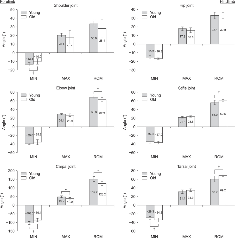
Fig. 3 Comparative depiction of the standardized minimum joint angle in degrees (MIN; maximum flexion), maximum joint angle in degrees (MAX; maximum extension) and range of motion in degrees (ROM) of the various joints for young (n = 5) and old (n = 5) Beagle dogs. The means ± SD are depicted. *Statistically significant difference between the groups (p < 0.05). †Tendency toward statistically significant difference between the groups (p < 0.10).
Reference
-
1. Abdelhadi J, Wefstaedt P, Galindo-Zamora V, Anders A, Nolte I, Schilling N. Load redistribution in walking and trotting Beagles with induced forelimb lameness. Am J Vet Res. 2013; 74:34–39.
Article2. Agostinho FS, Rahal SC, Miqueleto NSML, Verdugo MR, Inamassu LR, El-Warrak AO. Kinematic analysis of Labrador Retrievers and Rottweilers trotting on a treadmill. Vet Comp Orthop Traumatol. 2011; 24:185–191.
Article3. Bellows J, Colitz CMH, Daristotle L, Ingram DK, Lepine A, Marks SL, Sanderson SL, Tomlinson J, Zhang J. Common physical and functional changes associated with aging in dogs. J Am Vet Med Assoc. 2015; 246:67–75.
Article4. Bennett RL, DeCamp CE, Flo GL, Hauptman JG, Stajich M. Kinematic gait analysis in dogs with hip dysplasia. Am J Vet Res. 1996; 57:966–971.5. Beraud R, Moreau M, Lussier B. Effect of exercise on kinetic gait analysis of dogs afflicted by osteoarthritis. Vet Comp Orthop Traumatol. 2010; 23:87–92.
Article6. Bockstahler B, Kräutler C, Holler P, Kotschwar A, Vobornik A, Peham C. Pelvic limb kinematics and surface electromyography of the vastus lateralis, biceps femoris, and gluteus medius muscle in dogs with hip osteoarthritis. Vet Surg. 2012; 41:54–62.
Article7. Bockstahler B, Müller M, Henninger W, Mayrhofer E, Peham C, Podbregar I. Kinetic and kinematic motion analysis of the forelimbs in sound Malinois dogs – sampling of basic values. Wien Tierarztl Monatsschr. 2008; 95:127–138.8. Bockstahler BA, Henninger W, Müller M, Mayrhofer E, Peham C, Podbregar I. Influence of borderline hip dysplasia on joint kinematics of clinically sound Belgian Shepherd dogs. Am J Vet Res. 2007; 68:271–276.
Article9. Bockstahler BA, Prickler B, Lewy E, Holler PJ, Vobornik A, Peham C. Hind limb kinematics during therapeutic exercises in dogs with osteoarthritis of the hip joints. Am J Vet Res. 2012; 73:1371–1376.
Article10. Böddeker J, Drüen S, Meyer-Lindenberg A, Fehr M, Nolte I, Wefstaedt P. Computer-assisted gait analysis of the dog: comparison of two surgical techniques for the ruptured cranial cruciate ligament. Vet Comp Orthop Traumatol. 2012; 25:11–21.
Article11. Budsberg SC, Jevens DJ, Brown J, Foutz TL, DeCamp CE, Reece L. Evaluation of limb symmetry indices, using ground reaction forces in healthy dogs. Am J Vet Res. 1993; 54:1569–1574.12. Budsberg SC, Verstraete MC, Soutas-Little RW. Force plate analysis of the walking gait in healthy dogs. Am J Vet Res. 1987; 48:915–918.13. Catavitello G, Ivanenko YP, Lacquaniti F. Planar covariation of hindlimb and forelimb elevation angles during terrestrial and aquatic locomotion of dogs. PLoS One. 2015; 10:e0133936.
Article14. Colborne GR, Walker AM, Tattersall AJ, Fuller CJ. Effect of trotting velocity on work patterns of the hind limbs of Greyhounds. Am J Vet Res. 2006; 67:1293–1298.
Article15. Deban SM, Schilling N, Carrier DR. Activity of extrinsic limb muscles in dogs at walk, trot and gallop. J Exp Biol. 2012; 215:287–300.
Article16. Drüen S, Böddeker J, Meyer-Lindenberg A, Fehr M, Nolte I, Wefstaedt P. Computer-based gait analysis of dogs: evaluation of kinetic and kinematic parameters after cemented and cementless total hip replacement. Vet Comp Orthop Traumatol. 2012; 25:375–384.
Article17. Egenvall A, Bonnett BN, Olson P, Hedhammar A. Gender, age, breed and distribution of morbidity and mortality in insured dogs in Sweden during 1995 and 1996. Vet Rec. 2000; 146:519–525.
Article18. Eward C, Gillette RL, Eward W. Effects of unilaterally restricted carpal range of motion on kinematic gait analysis of the dog. Vet Comp Orthop Traumatol. 2003; 16:158–163.
Article19. Fischer MS, Blickhan R. The tri-segmented limbs of therian mammals: kinematics, dynamics, and self-stabilization—a review. J Exp Zool A Comp Exp Biol. 2006; 305:935–952.
Article20. Francuski JV, Radovanović A, Andrić N, Krstić V, Bogdanović D, Hadžić V, Todorović V, Lazarević Macanović M, Sourice Petit S, Beck-Cormier S, Guicheux J, Gauthier O, Kovačević Filipović M. Age-related changes in the articular cartilage of the stifle joint in non-working and working German Shepherd dogs. J Comp Pathol. 2014; 151:363–374.
Article21. Galindo-Zamora V, Dziallas P, Wolf DC, Kramer S, Abdelhadi J, Lucas K, Nolte I, Wefstaedt P. Evaluation of thoracic limb loads, elbow movement, and morphology in dogs before and after arthroscopic management of unilateral medial coronoid process disease. Vet Surg. 2014; 43:819–828.
Article22. Gillette RL, Craig Angle T. Recent developments in canine locomotor analysis: a review. Vet J. 2008; 178:165–176.
Article23. Gillette RL, Zebas CJ. A two-dimensional analysis of limb symmetry in the trot of Labrador retrievers. J Am Anim Hosp Assoc. 1999; 35:515–520.
Article24. Goldner B, Fuchs A, Nolte I, Schilling N. Kinematic adaptations to tripedal locomotion in dogs. Vet J. 2015; 204:192–200.
Article25. Helmsmüller D, Anders A, Nolte I, Schilling N. Ontogenetic change of the weight support pattern in growing dogs. J Exp Zool A Ecol Genet Physiol. 2014; 321:254–264.
Article26. Herzog W, Nigg BM, Read LJ, Olsson E. Asymmetries in ground reaction force patterns in normal human gait. Med Sci Sports Exerc. 1989; 21:110–114.
Article27. Hoskins JD. Preface. Geriatrics and Gerontology of the Dog and Cat. 2nd ed. St. Louis: Saunders;2004.28. Hottinger HA, DeCamp CE, Olivier NB, Hauptman JG, Soutas-Little RW. Noninvasive kinematic analysis of the walk in healthy large-breed dogs. Am J Vet Res. 1996; 57:381–388.29. Hutchinson D, Sutherland-Smith J, Watson AL, Freeman LM. Assessment of methods of evaluating sarcopenia in old dogs. Am J Vet Res. 2012; 73:1794–1800.
Article30. Jarvis SL, Worley DR, Hogy SM, Hill AE, Haussler KK, Reiser RF 2nd. Kinematic and kinetic analysis of dogs during trotting after amputation of a thoracic limb. Am J Vet Res. 2013; 74:1155–1163.
Article31. Kadaba MP, Ramakrishnan HK, Wootten ME, Gainey J, Gorton G, Cochran GVB. Repeatability of kinematic, kinetic, and electromyographic data in normal adult gait. J Orthop Res. 1989; 7:849–860.
Article32. Kim SE, Jones SC, Lewis DD, Banks SA, Conrad BP, Tremolada G, Abbasi AZ, Coggeshall JD, Pozzi A. In-vivo three-dimensional knee kinematics during daily activities in dogs. J Orthop Res. 2015; 33:1603–1610.
Article33. Kraft W. [Introduction]. [Geriatrics in Dogs and Cats]. 2nd ed. German: Parey Verlag, Stuttgart;2003. p. 23–31.34. Michell AR. Longevity of British breeds of dog and its relationships with sex, size, cardiovascular variables and disease. Vet Rec. 1999; 145:625–629.
Article35. Morgan JP, Pool RR, Miyabayashi T. Primary degenerative joint disease of the shoulder in a colony of beagles. J Am Vet Med Assoc. 1987; 190:531–540.36. Off W, Matis U. [Gait analysis in dogs. Part 2: Installation of a gait analysis laboratory and locomotor studies]. Tierarztl Prax. 1997; 25:303–311. German.37. Pagano TB, Wojcik S, Costagliola A, De Biase D, Iovino S, Iovane V, Russo V, Papparella S, Paciello O. Age related skeletal muscle atrophy and upregulation of autophagy in dogs. Vet J. 2015; 206:54–60.
Article38. Quinn MM, Keuler NS, Lu Y, Faria MLE, Muir P, Markel MD. Evaluation of agreement between numerical rating scales, visual analogue scoring scales, and force plate gait analysis in dogs. Vet Surg. 2007; 36:360–367.
Article39. Schofield JD, Weightman B. New knowledge of connective tissue ageing. J Clin Pathol Suppl (R Coll Pathol). 1978; 12:174–190.
Article40. Schwencke M, Smolders LA, Bergknut N, Gustås P, Meij BP, Hazewinkel HA. Soft tissue artifact in canine kinematic gait analysis. Vet Surg. 2012; 41:829–837.
Article41. Steiss JE, Yuill GT, White NA, Bowen JM. Modifications of a force plate system for equine gait analysis. Am J Vet Res. 1982; 43:538–540.42. Tian W, Cong Q, Menon C. Investigation on walking and pacing stability of german Shepherd dog for different locomotion speeds. J Bionic Eng. 2011; 8:18–24.
Article43. Torres BT, Whitlock D, Reynolds LR, Fu YC, Navik JA, Speas AL, Sornborger A, Budsberg SC. The effect of marker location variability on noninvasive canine stifle kinematics. Vet Surg. 2011; 40:715–719.
Article44. Waxman AS, Robinson DA, Evans RB, Hulse DA, Innes JF, Conzemius MG. Relationship between objective and subjective assessment of limb function in normal dogs with an experimentally induced lameness. Vet Surg. 2008; 37:241–246.
Article
- Full Text Links
-
- Actions
-
Cited
- CITED
-
- Close
- Share
-
- Similar articles
-
- Kinematic Gait Analysis of Ramp Walking in Normal Adult
- Difference in Gait Characteristics During Attention-Demanding Tasks in Young and Elderly Adults
- Comparing Self-Selected Speed Walking of the Elderly With Self-Selected Slow, Moderate, and Fast Speed Walking of Young Adults
- Changes in Gait Pattern After Surgeries for Equinus Gait in Cerebral Palsy Spastic Hemiplegia
- Gait analysis in clinically healthy small to toy breed dogs using a pressure plate
