Extensive silicone lymphadenopathy after breast implant insertion mimicking malignant lymphadenopathy
- Affiliations
-
- 1Department of Pathology, Korea University Hospital, Korea University College of Medicine, Seoul, Korea.
- 2Department of Radiology, Korea University Hospital, Korea University College of Medicine, Seoul, Korea.
- 3Department of Plastic Surgery and Reconstructive Surgery, Korea University Hospital, Korea University College of Medicine, Seoul, Korea.
- 4Division of Breast and Endocrine Surgery, Department of Surgery, Korea University Hospital, Korea University College of Medicine, Seoul, Korea. jungspil@korea.ac.kr
- KMID: 2396875
- DOI: http://doi.org/10.4174/astr.2017.93.6.331
Abstract
- Silicone implants are widely used in aesthetic and reconstructive breast surgery. Silicone lymphadenopathy is a well-known rare complication of implant insertion. Silicone leakage from a rupture or silicone bleeding can accumulate in lymph nodes. Foreign body reactions in the affected lymph nodes may be misdiagnosed as metastasis or malignant lymphadeno pathy upon initial presentation if silicone lymphadenopathy is not considered in the initial diagnosis. We report a case of siliconoma with extensive involvement of multiple lymph nodes mimicking malignant features to emphasize that clinicians should carefully evaluate each patient's medical history and disease status during differential diagnosis.
Keyword
MeSH Terms
Figure
-

Fig. 1 Ultrasonographic and computed tomographic findings of the lymph nodes. (A) Ultrasonographic images of axillary lymph nodes. Multiple hyperechoic lesions with diffuse posterior white noise (where silicone blocks sound transmission, white arrows) were observed. (B) Coronal view of computed tomography. Round, nonenhanced enlarged lymph nodes are located in the right axilla and neck (white arrows).
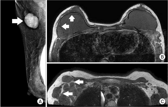
Fig. 2 Mammographic and MRI of the breast and axilla. (A) Mediola teral view of mam mo graphy im ages demon strating the right breast implant and an enlarged lymph node with the dense internal material in the right axilla (white arrow). (B) Axial nonfat sup pressed, T1-weighted MRI of the breast show tear drop sign within the right implant and a hypo dense thin line in the interior of the implant indicative of intracapsular rupture (white arrows). (C) Axial nonfat suppressed, T1-weighted MRI im ages of the breast show oval cir cumscribed lymph nodes with isosignal intensity (white arrows) in the right axilla compared to the implanted silicone bag in panel B.
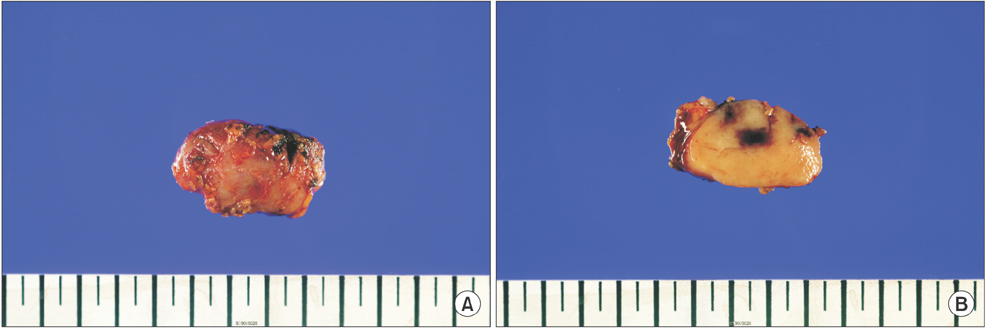
Fig. 3 Macroscopic findings of the excised lymph node. (A) The exterior of the lymph node is round and reddish-brown in color. (B) The cut surface of the node.
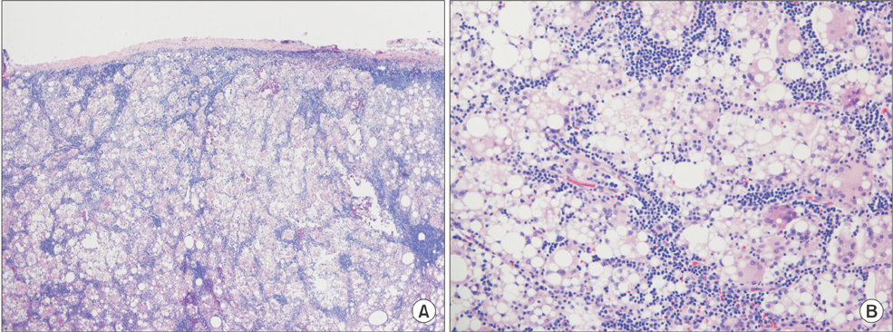
Fig. 4 Microscopic findings of excised lymph node. (A) Silicone-containing histiocytes are scattered through the lymph node parenchyme (H&E, ×40). (B) Large clusters of silicone-filled, clear-appearing histiocytes have replaced lymphoid tissue (H&E, ×200).
Reference
-
1. Gidengil CA, Predmore Z, Mattke S, van Busum K, Kim B. Breast implant-associated anaplastic large cell lymphoma: a systematic review. Plast Reconstr Surg. 2015; 135:713–720.2. Cook PD, Osborne BM, Connor RL, Strauss JF. Follicular lymphoma adja cent to foreign body granulomatous inflammation and fibrosis surrounding silicone breast prosthesis. Am J Surg Pathol. 1995; 19:712–717.3. Gundeslioglu AO, Hakverdi S, Erdem O, Ozen EC, Inan I, Emlik D. Axillary lipogranuloma mimicking carcinoma metastasis after silicone breast implant rupture: a case report. J Plast Reconstr Aesthet Surg. 2013; 66:e72–e75.4. Maxwell GP, Gabriel A. The evolution of breast implants. Clin Plast Surg. 2015; 36:399–404.5. McLaughlin JK, Lipworth L, Murphy DK, Walker PS. The safety of silicone gelfilled breast implants: a review of the epidemiologic evidence. Ann Plast Surg. 2007; 59:569–580.6. de Jong D, Vasmel WL, de Boer JP, Verhave G, Barbe E, Casparie MK, et al. Anaplastic largecell lymphoma in women with breast implants. JAMA. 2008; 300:2030–2035.7. Holmich LR, Friis S, Fryzek JP, Vejborg IM, Conrad C, Sletting S, et al. Incidence of silicone breast implant rupture. Arch Surg. 2003; 138:801–806.8. Allergan Silicone Breast Implant U.S. Core Clinical Study Group. Spear SL, Murphy DK. Natrelle round silicone breast implants: Core Study results at 10 years. Plast Reconstr Surg. 2014; 133:1354–1361.9. Zambacos GJ, Molnar C, Mandrekas AD. Silicone lymphadenopathy after breast augmentation: case reports, review of the literature, and current thoughts. Aesthetic Plast Surg. 2013; 37:278–289.10. Buley ID. Fine needle aspiration of lymph nodes. J Clin Pathol. 1998; 51:881–885.
- Full Text Links
-
- Actions
-
Cited
- CITED
-
- Close
- Share
-
- Similar articles
-
- Axillary silicone lymphadenopathy caused by gel bleeding with intact silicone breast implants: a case report
- The Clinical Implications of Poly Implant Prothese Breast Implants: An Overview
- Silicone Granuloma Mimicking a Lymphatic Metastasis in a Lung Cancer Patient: A Case Report
- Understanding Silicone Breast Implant-Associated Complications for Radiologists
- Toxocariasis Mimicking Lymphoma and Presenting as Multiple Lymphadenopathy: A Case Report
