Clin Exp Otorhinolaryngol.
2017 Dec;10(4):338-343. 10.21053/ceo.2017.00080.
Radiographic Changes of the Nasal Septal Body Among Patients With Sinonasal Diseases
- Affiliations
-
- 1Department of Otorhinolaryngology-Head and Neck Surgery, Seoul National University Bundang Hospital, Seoul National University College of Medicine, Seoul, Korea. csrhee@snu.ac.kr
- 2Department of Otorhinolaryngology-Head and Neck Surgery, University of Santo Tomas Hospital, Manila, Philippines.
- 3Research Center for Sensory Organs, Seoul National University Medical Research Center, Seoul, Korea.
- 4Institute of Allergy and Clinical Immunology, Seoul National University Medical Research Center, Seoul, Korea.
- KMID: 2396789
- DOI: http://doi.org/10.21053/ceo.2017.00080
Abstract
OBJECTIVES
This study aims to determine the anatomical changes occurring in the nasal septal body (NSB) among patients with sinonasal disease and compares the measurements obtained from patients without sinonasal disease.
METHODS
This was a retrospective study involving review of 405 (93 without and 212 with sinonasal disease) plain paranasal sinus computed tomography (PNS CT) on coronal view at a tertiary training hospital, which was done consecutively from January 2014 to December 2015. NSB measurements from 3 areas were done: anterior part (A), located anterior and superior to inferior turbinate; middle or widest (M) part, located anterior to middle turbinate and superior to inferior turbinate and posterior (P) part, located within the anterior 1/3 of middle turbinate not going beyond the crista galli. Posterior part of septum (sP) was measured at the area of horizontal attachment of middle turbinate to the lateral nasal wall and superior turbinate to represent the less vasoactive part of the septum. Demographic data and NSB diameters were also analyzed.
RESULTS
The mean NSB diameter measurements were significantly larger among the diseased group (disease vs. control; A: 6.88 mm vs. 5.92 mm, P=0.001; M: 12.74 mm vs. 10.47 mm, P=0.001; P: 8.35 mm vs. 6.79 mm, P=0.001). A similar observation in sP part (3.35 mm vs. 2.54 mm, P=0.014) was noted. When grouped by age, among the control group, older subjects had considerable decrease in NSB size in all points of measurements except for sP (P>0.05). Such reduction in size is noticeable for those in their 4th, 5th, 6th, and 7th decades of life. For the diseased group, a trend of increasing NSB and sP size was noted as the subjects are getting older. However, only the anterior part (A) of NSB reached statistical significance (P=0.016).
CONCLUSION
With aging we expect nasal mucosal atrophy among normal subjects. However, patients with chronic sinonasal disease showed thickened nasal mucosa. Further study for the reversibility of thickened mucosa is needed.
Keyword
Figure
-
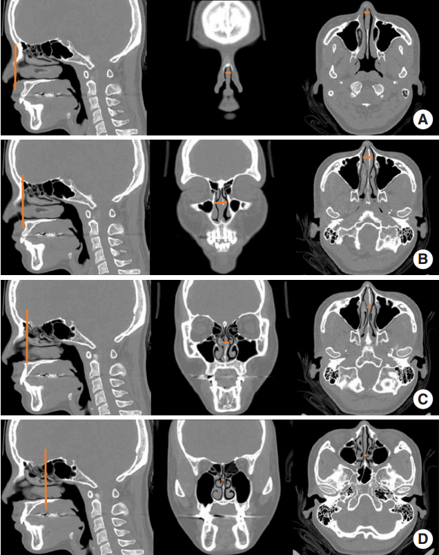
Fig. 1. Measurement of nasal septal body (NSB) and posterior sep tum. The red lines in left side of figures indicate the level of measurement, and those in middle and right sides of figures indicate the actual measurement. (A) NSB anterior part, (B) NSB middle or widest part, (C) NSB posterior part, and (D) posterior part of septum.
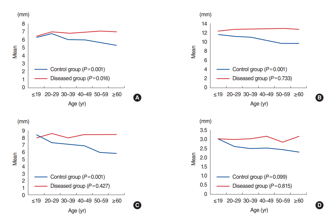
Fig. 2. Nasal septal body (NSB) diameter by age group between control and diseased group. (A) NSB anterior part, (B) NSB middle or widest part, (C) NSB posterior part, and (D) posterior part of septum.
Reference
-
1. Costa DJ, Sanford T, Janney C, Cooper M, Sindwani R. Radiographic and anatomic characterization of the nasal septal swell body. Arch Otolaryngol Head Neck Surg. 2010; Nov. 136(11):1107–10.
Article2. Wexler D, Braverman I, Amar M. Histology of the nasal septal swell body (septal turbinate). Otolaryngol Head Neck Surg. 2006; Apr. 134(4):596–600.
Article3. Setlur J, Goyal P. Relationship between septal body size and septal deviation. Am J Rhinol Allergy. 2011; Nov-Dec. 25(6):397–400.
Article4. Elwany S, Salam SA, Soliman A, Medanni A, Talaat E. The septal body revisited. J Laryngol Otol. 2009; Mar. 123(3):303–8.
Article5. Cole P. The four components of the nasal valve. Am J Rhinol. 2003; Mar-Apr. 17(2):107–10.
Article6. Farmer SE, Eccles R. Chronic inferior turbinate enlargement and the implications for surgical intervention. Rhinology. 2006; Dec. 44(4):234–8.7. Arslan M, Muderris T, Muderris S. Radiological study of the intumescentia septi nasi anterior. J Laryngol Otol. 2004; Mar. 118(3):199–201.
Article8. San T, Gurkan E, Erdogan B, Tasel B. The effect of allergic rhinitis on nasal septal body size. J Med Updates. 2014; 4(2):49–51.
Article9. Brozek JL, Bousquet J, Baena-Cagnani CE, Bonini S, Canonica GW, Casale TB, et al. Allergic Rhinitis and its Impact on Asthma (ARIA) guidelines: 2010 revision. J Allergy Clin Immunol. 2010; Sep. 126(3):466–76.10. Kim DK, Rhee CS, Han DH, Won TB, Kim DY, Kim JW. Treatment of allergic rhinitis is associated with improved attention performance in children: the Allergic Rhinitis Cohort Study for Kids (ARCO-Kids). PLoS One. 2014; Oct. 9(10):e109145.
Article11. Rosenfeld RM, Piccirillo JF, Chandrasekhar SS, Brook I, Ashok Kumar K, Kramper M, et al. Clinical practice guideline (update): adult sinusitis. Otolaryngol Head Neck Surg. 2015; Apr. 152(2 Suppl):S1–S39.
Article12. Ashraf N, Bhattacharyya N. Determination of the “incidental” Lund score for the staging of chronic rhinosinusitis. Otolaryngol Head Neck Surg. 2001; Nov. 125(5):483–6.
Article13. Miman MC, Deliktas H, Ozturan O, Toplu Y, Akarcay M. Internal nasal valve: revisited with objective facts. Otolaryngol Head Neck Surg. 2006; Jan. 134(1):41–7.
Article14. Pinto JM, Jeswani S. Rhinitis in the geriatric population. Allergy Asthma Clin Immunol. 2010; May. 6(1):10.
Article15. Rehl RM, Balla AA, Cabay RJ, Hearp ML, Pytynia KB, Joe SA. Mucosal remodeling in chronic rhinosinusitis. Am J Rhinol. 2007; Nov-Dec. 21(6):651–7.
Article16. Chan KH, Abzug MJ, Coffinet L, Simoes EA, Cool C, Liu AH. Chronic rhinosinusitis in young children differs from adults: a histopathology study. J Pediatr. 2004; Feb. 144(2):206–12.
Article17. Pawankar R, Nonaka M. Inflammatory mechanisms and remodeling in chronic rhinosinusitis and nasal polyps. Curr Allergy Asthma Rep. 2007; Jun. 7(3):202–8.
Article18. Wotman M, Kacker A. Should otolaryngologists pay more attention to nasal swell bodies? Laryngoscope. 2015; Aug. 125(8):1759–60.
Article19. Haight JS, Gardiner GW. Nasal cryosurgery and cautery: should the septum be treated and is a diagnosis relevant? J Otolaryngol. 1989; Jun. 18(4):144–50.20. Yu MS, Kim JY, Kim BH, Kang SH, Lim DJ. Feasibility of septal body volume reduction for patients with nasal obstruction. Laryngoscope. 2015; Jul. 125(7):1523–8.
Article21. Catalano P, Ashmead MG, Carlson D. Radiofrequency ablation of septal swell body. Ann Otolaryngol Rhinol. 2015; Nov. 2(11):1069.
- Full Text Links
-
- Actions
-
Cited
- CITED
-
- Close
- Share
-
- Similar articles
-
- Clinical Analysis of Benign and Malignant Nasal Septal Tumors
- Nasal Septal Swell Body: Radiologic Characteristics and the Relationship to Nasal Septal Deviation
- Nasal Septal Perforation due to Button Battery
- Structural Changes of Inferior Turbinate in Patients with Septal Deviation: Surgical Implication
- Using nasal septal cartilage-bone complex for anterior septal reconstruction in Asians
