Korean J Ophthalmol.
2017 Dec;31(6):572-573. 10.3341/kjo.2017.0103.
Spontaneous Resolution of Macular Hole with Retinal Detachment in a Highly Myopic Eye
- Affiliations
-
- 1Department of Ophthalmology, Dongsan Medical Center, Keimyung University School of Medicine, Daegu, Korea. eyedr@dsmc.or.kr
- KMID: 2396710
- DOI: http://doi.org/10.3341/kjo.2017.0103
Abstract
- No abstract available.
Figure
-
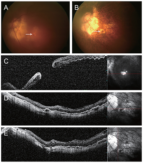
Fig. 1 Fundus photographs and optical coherence tomography. (A) Fundus photograph of macular hole (arrow) with retinal detachment. (B) After 2 years, spontaneous resolution of macular hole (arrow) and associated retinal detachment were observed. Atrophic scar change in the fovea was seen on fundus photographs. (C) Macular hole with retinal detachment. (D,E) After 2 years, complete retinal reattachment was observed, without any retinoschisis or macular hole on horizontal- and vertical-view optical coherence tomography.
Reference
-
1. Alkabes M, Pichi F, Nucci P, et al. Anatomical and visual outcomes in high myopic macular hole (HM-MH) without retinal detachment: a review. Graefes Arch Clin Exp Ophthalmol. 2014; 252:191–199.2. Min WK. Spontaneous reattachment of retinal detachment with macular hole in nonmyopic patients. Korean J Ophthalmol. 1995; 9:66–68.3. Tam BS, Kwok AK, Bhende P, Lam DS. Spontaneous reattachment of retinal detachment in a highly myopic eye with a macular hole. Eye (Lond). 2000; 14(Pt 4):661–662.4. Li Y, Jonas JB, Lu L. Spontaneous closure of highly myopic macular hole associated with retinal detachment. Acta Ophthalmol. 2014; 92:e408–e410.5. Yu J, Jiang C, Xu G. Spontaneous closure of a myopic macular hole with retinal reattachment in an eye with high myopia and staphyloma: a case report. BMC Ophthalmol. 2014; 14:111.
- Full Text Links
-
- Actions
-
Cited
- CITED
-
- Close
- Share
-
- Similar articles
-
- Spontaneous reattachment of retinal detachment with macular hole in nonmyopic patients
- Primary Silicone Oil Tamponade with Vitrectomy in Macular Hole Retinal Detachment of Highly Myopic Eyes
- Three Cases of Macular Buckling for Retinal Detachment due to Macular Hole in Highly Myopic Eyes
- Effect of Internal Limiting Membrane Removal in Treatment of Retinal Detachment Caused by Myopic Macular Hole
- Treatment of Macular Hole Retinal Detachment
