Yonsei Med J.
2009 Apr;50(2):222-226.
External Jugular Vein Catheterization Using 'Intra-Atrial Electrocardiogram'
- Affiliations
-
- 1Department of Anesthesiology and Reanimation, Suleyman Demirel University School of Medicine, Isparta, Turkey. dilekkaraaslan1@yahoo.com
Abstract
- PURPOSE
To investigate the reliability of intra-atrial electrocardiogram (ECG) use for external jugular vein (EJV) catheterization. MATERIALS AND METHODS: Patients undergoing open heart surgery in Suleyman Demirel University Hospital between February and June 2006 were included in the study. Using a sterile Seldinger technique, a triple lumen polyurethane central venous catheter was introduced (Certofix(R) Trio V 720, length 20 cm, 7 French) under intra-atrial ECG guidance. The presence of an increase in P-wave size was recorded. Just after the surgery, a portable chest X-ray was taken. The method was considered to be successful when a change in P-wave could be seen and the catheter was in the superior vena cava, as well as when there was no change in P-wave and the catheter was not in the superior vena cava. RESULTS: In six patients (12%), we were not able to advance the guidewire. In the remaining 44 patients, the catheter was inserted without problem. Eight of these 44 catheters were positioned in the innominate vein, with a malposition ratio of 18%. The success rate of external jugular vein cannulation with intra-atrial ECG was 95%. No complications occured related to the EJV cannulation. CONCLUSION: Considering that it is easily accessed without complication, and the malposition is successfully detected by intra-atrial ECG, EJV is a suitable access for central venous cannulation when internal jugular vein (IJV) is not usable.
MeSH Terms
Figure
-
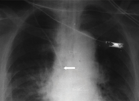
Fig. 1 Appropriate localization of the external jugular vein catheter on chest X-ray. Catheter tip marked with an arrow.

Fig. 2 Fluoroscopic view of the external jugular vein catheter placed in the left innominate vein. Catheter tip marked with an arrow.
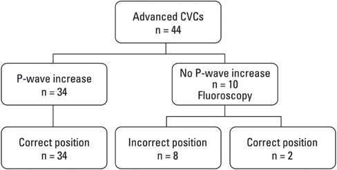
Fig. 3 Evaluation of the method.
Reference
-
1. Weeks SM. Unconventional venous access. Tech Vasc Interv Radiol. 2002. 5:114–120.
Article2. Trerotola SO, Kuhn-Fulton J, Johnson MS, Shah H, Ambrosius WT, Kneebone PH. Tunneled infusion catheters: increased incidence of symptomatic venous thrombosis after subclavian versus internal jugular venous access. Radiology. 2000. 217:89–93.
Article3. Trerotola SO. You are asked to place a dialysis access catheter in a patient. What is your preferred access site, and why? J Vasc Interv Radiol. 1997. 8:75–76.4. Macdonald S, Watt AJ, McNally D, Edwards RD, Moss JG. Comparison of technical success and outcome of tunneled catheters inserted via the jugular and subclavian approaches. J Vasc Interv Radiol. 2000. 11:225–231.
Article5. Schillinger F, Schillinger D, Montagnac R, Milcent T. Post catheterisation vein stenosis in haemodialysis: comparative angiographic study of 50 subclavian and 50 internal jugular accesses. Nephrol Dial Transplant. 1991. 6:722–724.
Article6. Moss AH, McLaughlin MM, Lempert KD, Holley JL. Use of a silicone catheter with a Dacron cuff for dialysis short-term vascular access. Am J Kidney Dis. 1988. 12:492–498.
Article7. Cho SK, Shin SW, Do YS, Park KB, Choo SW, Choo IW. Use of the right external jugular vein as the preferred access site when the right internal jugular vein is not usable. J Vasc Interv Radiol. 2006. 17:823–829.
Article8. Nicolson SC, Sweeney MF, Moore RA, Jobes DR. Comparison of internal and external jugular cannulation of central circulation in the pediatric patient. Crit Care Med. 1985. 13:747–749.
Article9. Chu KS, Hsu JH, Wang SS, Tang CS, Cheng KI, Wang CK, et al. Accurate central venous port-A catheter placement: intravenous electrocardiography and surface landmark techniques compared by using transesophageal echocardiography. Anesth Analg. 2004. 98:910–914.
Article10. Watters VA, Grant JP. Use of electrocardiogram to position right atrial catheters during surgery. Ann Surg. 1997. 225:165–171.
Article11. Schummer W, Herrmann S, Schummer C, Funke F, Steenbeck J, Fuchs J, et al. Intra-atrial ECG is not a reliable method for positioning left internal jugular vein catheters. Br J Anaesth. 2003. 91:481–486.
Article12. Mickley V. Central venous catheters: many questions, few answers. Nephrol Dial Transplant. 2002. 17:1368–1373.
Article13. McCowan TC, Ferris EJ, Carver DK, Harshfield DL. Use of external jugular vein as a route for percutaneous inferior vena caval filter placement. Radiology. 1990. 176:527–530.
Article14. Stickle BR, McFarlane H. Prediction of a small internal jugular vein by external jugular vein diameter. Anaesthesia. 1997. 52:220–222.
Article15. Belani KG, Buckley JJ, Gordon JR, Castaneda W. Percutaneous cervical central venous line placement: a comparison of the internal and external jugular vein routes. Anesth Analg. 1980. 59:40–44.16. Jobes DR, Schwartz AJ, Greenhow DE, Stephenson LW, Ellison N. Safer jugular vein cannulation: recognition of arterial puncture and preferential use of the external jugular route. Anesthesiology. 1983. 59:353–355.17. Malatinsky J, Kadlic T, Májek M, Sámel M. Misplacement and loop formation of central venous catheters. Acta Anaesthesiol Scand. 1976. 20:237–247.
Article18. Salmela L, Aromaa U. Verification of the position of a central venous catheter by intra-atrial ECG. When does this method fail? Acta Anaesthesiol Scand. 1993. 37:26–28.
Article19. Illustrated encyclopedia of human anatomic variation [www document]. accessed on 25 May 2007. URL http://www.anatomyatlases.org/AnatomicVariants/AnatomyHP.shtml.20. McGee WT, Ackerman BL, Rouben LR, Prasad VM, Bandi V, Mallory DL. Accurate placement of central venous catheters: a prospective, randomized, multicenter trial. Crit Care Med. 1993. 21:1118–1123.21. Vesely TM. Central venous catheter tip position: a continuing controversy. J Vasc Interv Radiol. 2003. 14:527–534.
Article22. Fletcher SJ, Bodenham AR. Safe placement of central venous catheters: where should the tip of the catheter lie? Br J Anaesth. 2000. 85:188–191.23. Peres PW. Positioning central venous catheters-a prospective survey. Anaesth Intensive Care. 1990. 18:536–539.24. Aslamy Z, Dewald CL, Heffner JE. MRI of central venous anatomy: implications for central venous catheter insertion. Chest. 1998. 114:820–826.25. Hsu JH, Wang CK, Chu KS, Cheng KI, Chuang HY, Jaw TS, et al. Comparison of radiographic landmarks and the echocardiographic SVC/RA junction in the positioning of long-term central venous catheters. Acta Anaesthesiol Scand. 2006. 50:731–735.
Article26. Schummer W, Schummer C, Schelenz C, Brandes H, Stock U, Müller T, et al. Central venous catheters-the inability of 'intra--atrial ECG' to prove adequate positioning. Br J Anaesth. 2004. 93:193–198.
Article
- Full Text Links
-
- Actions
-
Cited
- CITED
-
- Close
- Share
-
- Similar articles
-
- A Large Subcutaneous Hematoma during Internal Jugular Vein Catheterization: A case report
- Roentgenographic Confirmation of Central Venous Catheter Tips through the External and Internal Jugular Veins in Children
- Roentgenographic Confirmation of Central Venous Catheter Tips Through the Basilic and External Jugular Veins
- Evaluation of the Technique of Central Venous Catheterization via the External Jugular Vein
- Internal jugular vein thrombosis detection by ultrasound scan after failure of internal jugular vein catheterization: A case report
