Yonsei Med J.
2009 Apr;50(2):211-214.
Higher Lesion Detection by 3.0T MRI in Patient with Transient Global Amnesia
- Affiliations
-
- 1Department of Neurology, Yonsei University College of Medicine, Seoul, Korea. kylee@yuhs.ac
- 2Department of Radiology, Yonsei University College of Medicine, Seoul, Korea.
Abstract
- PURPOSE
Transient global amnesia (TGA) patients were retrospectively reviewed to determine the usefulness of high-field strength MRI in detecting probable ischemic lesions in TGA. MATERIALS AND METHODS: We investigated the lesion detection rate in patients with TGA using 1.5T and 3.0T MRI. Acute probable ischemic lesions were defined as regions of high-signal intensity in diffusion weighted image with corresponding low-signal intensity in apparent diffusion coefficient map. RESULTS: 3.0T MRI showed 11 out of 32 patients with probable ischemic lesions in the hippocampus with mean lesion size of 2.8 +/- 0.6 mm, whereas 1.5T MRI detected no lesion in any of 11 patients. There were no significant differences in clinical characteristics between the groups of 1.5 and 3.0T MRI. CONCLUSION: High-field strength MRI has a higher detection rate of probable ischemic lesions than low-field strength MRI in patients with TGA.
Keyword
MeSH Terms
Figure
-
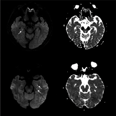
Fig. 1 Representative 3.0T MRI of 2 TGA patients shows small high-signal intensity lesions in the hippocampus on DWI (arrow) and corresponding low-signal intensity lesions on ADC map (arrow head). TGA, transient global amnesia; DWI, diffusion weighted imaging; ADC, apparent diffusion coefficient.
Reference
-
1. Hodges JR, Warlow CP. Syndromes of transient amnesia: towards a classification. A study of 153 cases. J Neurol Neurosurg Psychiatry. 1990. 53:834–843.
Article2. Sander K, Sander D. New insights into transient global amnesia: recent imaging and clinical findings. Lancet Neurol. 2005. 4:437–444.
Article3. Sedlaczek O, Hirsch JG, Grips E, Peters CN, Gass A, Wöhrle J, et al. Detection of delayed focal MR changes in the lateral hippocampus in transient global amnesia. Neurology. 2004. 62:2165–2170.
Article4. Winbeck K, Etgen T, von Einsiedel HG, Röttinger M, Sander D. DWI in transient global amnesia and TIA: proposal for an ischaemic origin of TGA. J Neurol Neurosurg Psychiatry. 2005. 76:438–441.
Article5. Felix MM, Castro LH, Maia AC Jr, da Rocha AJ. Evidence of acute ischemic tissue change in transient global amnesia in magnetic resonance imaging: case report and literature review. J Neuroimaging. 2005. 15:203–205.
Article6. Kapur N. Transient epileptic amnesia--a clinical update and a reformulation. J Neurol Neurosurg Psychiatry. 1993. 56:1184–1190.
Article7. Lewis SL. Aetiology of transient global amnesia. Lancet. 1998. 352:397–399.
Article8. Inzitari D, Pantoni L, Lamassa M, Pallanti S, Pracucci G, Marini P. Emotional arousal and phobia in transient global amnesia. Arch Neurol. 1997. 54:866–873.
Article9. Huber R, Aschoff AJ, Ludolph AC, Riepe MW. Transient Global Amnesia. Evidence against vascular ischemic etiology from diffusion weighted imaging. J Neurol. 2002. 249:1520–1524.10. Gass A, Gaa J, Hirsch J, Schwartz A, Hennerici MG. Lack of evidence of acute ischemic tissue change in transient global amnesia on single-shot echo-planar diffusion-weighted MRI. Stroke. 1999. 30:2070–2072.
Article11. Lee HY, Kim JH, Weon YC, Lee JS, Kim SY, Youn SW, et al. Diffusion-weighted imaging in transient global amnesia exposes the CA1 region of the hippocampus. Neuroradiology. 2007. 49:481–487.
Article12. Toledo M, Pujadas F, Grivé E, Alvarez-Sabin J, Quintana M, Rovira A. Lack of evidence for arterial ischemia in transient global amnesia. Stroke. 2008. 39:476–479.
Article13. Willinek WA, Schild HH. Clinical advantages of 3.0 T MRI over 1.5 T. Eur J Radiol. 2008. 65:2–14.14. Kuhl CK, Textor J, Gieseke J, von Falkenhausen M, Gernert S, Urbach H, et al. Acute and subacute ischemic stroke at high-field-strength (3.0-T) diffusion-weighted MR imaging: intraindividual comparative study. Radiology. 2005. 234:509–516.
Article15. Benameur K, Bykowski JL, Luby M, Warach S, Latour LL. Higher prevalence of cortical lesions observed in patients with acute stroke using high-resolution diffusion-weighted imaging. AJNR Am J Neuroradiol. 2006. 27:1987–1989.16. Bertrand A, Oppenheim C, Lamy C, Rodrigo S, Naggara O, Mas JL, et al. Comparison of optimized and standard diffusion-weighted imaging at 1.5T for the detection of acute lesions in patients with transient ischemic attack. AJNR Am J Neuroradiol. 2008. 29:363–365.
Article17. Bartsch T, Alfke K, Stingele R, Rohr A, Freitag-Wolf S, Jansen O, et al. Selective affection of hippocampal CA-1 neurons in patients with transient global amnesia without long-term sequelae. Brain. 2006. 129:2874–2884.
Article
- Full Text Links
-
- Actions
-
Cited
- CITED
-
- Close
- Share
-
- Similar articles
-
- Recurrent transient amnesia: a case of transient epileptic amnesia misdiag-nosed as transient global amnesia
- Retrograde Amnesia as a Predominant Symptom of Transient Global Amnesia
- Transient Global Amnesia Developed in a 14-Year Old Girl during Cellular Phone Texting
- Transient Global Amnesia after Gastroscopy
- A Clinical Study of Transient Global Amnesia
