Treatment of Melasma and Post-Inflammatory Hyperpigmentation by a Picosecond 755-nm Alexandrite Laser in Asian Patients
- Affiliations
-
- 1Department of Dermatology, Asan Medical Center, University of Ulsan College of Medicine, Seoul, Korea. changse2016@gmail.com
- 2Chois Dermatology Clinic, Seoul, Korea.
- KMID: 2395188
- DOI: http://doi.org/10.5021/ad.2017.29.6.779
Abstract
- The picosecond lasers have shown to effectively treat tattoo pigments that are intractable to previous multiple Q-switched (QS) laser treatments. Therefore we hypothesized that a picosecond laser would show better efficacy with minimal adverse events in the treatment of melasma and post-inflammatory hyperpigmentation (PIH) that are difficult to treat with conventional QS lasers. Two patients with melasma and one patient with PIH were treated with a Picosecond 755-nm Alexandrite Laser (Cyanosure, USA). All patients were Korean with skin type IV and no longer responding to QS laser treatments. Laser treatment was well tolerated in all the patients. Adverse events such as PIH were not reported during 8 weeks of follow up period. After the multiple treatment sessions, one patient reported fair improvement and two patients reported good improvement. Consistent with the clinical results, ex vivo skin model irradiated with a Picosecond 755-nm Alexandrite Laser also showed decreased epidermal keratinocyte necrosis compared with the 532-nm QS Neodymium-Doped Yttrium Aluminium Garnet Laser (Lutronic, Korea) yet decreased melanin content. In conclusion, the Picosecond 755-nm Alexandrite Laser may be useful for effective treatment of intractable melasma and PIH with fewer adverse events in dark Asian skin.
MeSH Terms
Figure
-
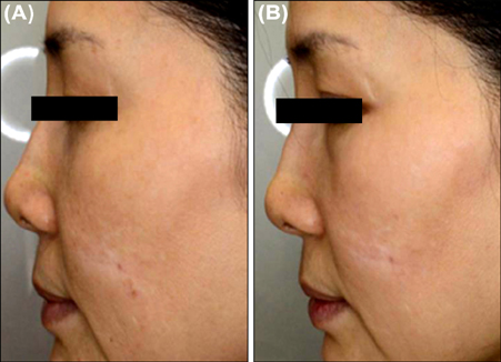
Fig. 1 Melasma in a 53-year-old female. (A) Findings at baseline. (B) Findings after six treatments of 0.57 J/cm2 with a 6-mm spot size using a Picosecond 755-nm Alexandrite Laser.

Fig. 2 Melasma in a 45-year-old female. (A) Findings at baseline. (B) Findings after 14 treatments of 0.57 J/cm2 with a 6-mm spot size using a Picosecond 755-nm Alexandrite Laser.
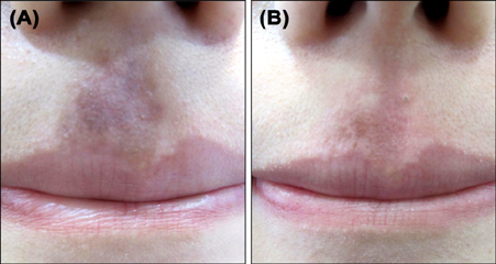
Fig. 3 Post-inflammatory hyperpigmentation in a 20-year-old female. (A) Findings at baseline. (B) Findings after seven treatments of 5.25 J/cm2 with a 2-mm spot size using a Picosecond 755-nm Alexandrite Laser.

Fig. 4 (A~D) Photomicrographs of a skin model stained for nitro blue tetrazolium after laser treatment: (A) control, (B) 1,064-nm Q-switched Neodymium-Doped Yttrium Aluminium Garnet (QSNY) Laser, (C) 532-nm QSNY Laser, (D) Picosecond 755-nm Alexandrite Laser; A~D, ×200). (E) Relative value of melanin index seven days after treatment. Quantitative measurement of melanin pigments was performed using Image J analysis following Fontana-Masson staining.
Reference
-
1. Kim BW, Lee MH, Chang SE, Yun WJ, Won CH, Lee MW, et al. Clinical efficacy of the dual-pulsed Q-switched neodymium:yttrium-aluminum-garnet laser: comparison with conservative mode. J Cosmet Laser Ther. 2013; 15:340–341.
Article2. Ross V, Naseef G, Lin G, Kelly M, Michaud N, Flotte TJ, et al. Comparison of responses of tattoos to picosecond and nanosecond Q-switched neodymium: YAG lasers. Arch Dermatol. 1998; 134:167–171.
Article3. Saedi N, Metelitsa A, Petrell K, Arndt KA, Dover JS. Treatment of tattoos with a picosecond alexandrite laser: a prospective trial. Arch Dermatol. 2012; 148:1360–1363.
Article4. Chesnut C, Diehl J, Lask G. Treatment of nevus of ota with a picosecond 755-nm alexandrite laser. Dermatol Surg. 2015; 41:508–510.
Article5. Kim JE, Chang SE, Yeo UC, Haw S, Kim IH. Histopathological study of the treatment of melasma lesions using a low-fluence Q-switched 1064-nm neodymium: yttrium-aluminium-garnet laser. Clin Exp Dermatol. 2013; 38:167–171.
Article
- Full Text Links
-
- Actions
-
Cited
- CITED
-
- Close
- Share
-
- Similar articles
-
- A Pilot Split-Neck Case Study to Compare the Efficacy of the Long-Pulsed 755 nm Laser and the 532 nm Picosecond Laser for Acrochordon Removal
- Picosecond laser treatment for Asian skin pigments: a review
- Effectiveness of 250-picosecond laser for the treatment of melasma and pigmented lesions with a low fluence 1,064-nm Nd:YAG laser in Republic of Korea: retrospective study
- A split-face study evaluating the efficacy of a topical antioxidant cream containing tocotrienol after 1064-nm picosecond Nd:YAG laser treatment for environment-induced skin pigmentation
- Acropigmentation Symmetrica of Dohi Treated with the Q-switched Alexandrite Laser
