Accompanying Pulmonary Arteriovenous Malformation in Patient with Hydatidiform Mole: A Case Report
- Affiliations
-
- 1Department of Radiology, Kangwon National University Hospital, Chuncheon, Korea. hk2005.yoon@gmail.com
- 2Department of Obstetrics and Gynecology, Kangdong Sacred Heart Hospital, Hallym University College of Medicine, Seoul, Korea.
- 3Division of Cardiology, Department of Internal Medicine, Kangwon National University School of Medicine, Kangwon National University Hospital, Chuncheon, Korea.
- KMID: 2394049
- DOI: http://doi.org/10.3348/jksr.2017.77.5.339
Abstract
- The most common site of metastasis in gestational trophoblastic disease (GTD) is the lung. To the best of our knowledge, arteriovenous malformations (AVMs) associated with pulmonary metastatic lesions are extremely rare in patients with GTD. Here, we report a case of a nineteen-year-old woman who presented with pulmonary AVMs which developed in the areas of pulmonary metastases, complicated by recent hemorrhage.
MeSH Terms
Figure
-
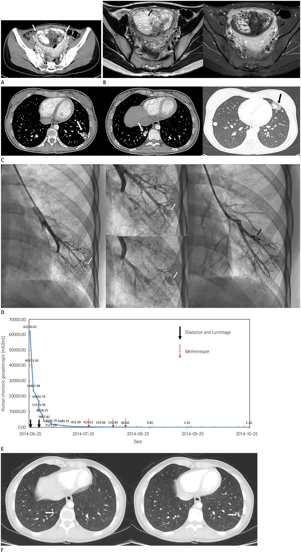
Fig. 1 A 19-year-old woman with hydatidiform mole, metastasizing to the lung accompanying with the pulmonary; arteriovenous malformation. A. Contrast-enhanced abdomen CT shows an enlarged uterus with abnormal heterogeneous enhancement of the endometrium (white thick arrows). Multiple air bubbles are found in the uterine cavity because dilatation and curettage is performed on the same day. B. Axial T2-weighted (left) and T1-weighted gadolinium-enhanced (right) pelvic MR images demonstrate strong enhancement (white thin arrows) of the endometrium with multiple T2-hyperintense cystic foci (a black thick arrow). C. Axial contrast-enhanced chest CT shows multiple nodules consistent with pulmonary metastases. Some nodules communicate with a curvilinear structure (white thick arrows), indicating arteriovenous malformations. Ground-glass halo around a metastatic nodule in the inferior lingular segment of the left upper lobe represents hemorrhage (a black arrow). D. Pulmonary angiography and coil embolization. Selective left pulmonary arteriogram shows pulmonary AVM (white thick arrows). Embolization coil (a black arrow) is deployed and postembolization pulmonary arteriogram shows complete occlusion of AVM. AVM = arteriovenous malformation, CT = computed tomography E. hCG levels during the course of treatment. At the time of diagnosis, the serum hCG level was 62,200.02 mIU/mL. The serum hCG level decreased to 3897.82 mIU/mL after two dilatation and curettage on June 26 and July 2, 2014. From July 28 to August 11, the patient was given three cycles of Methotrexate weekly. The serum hCG level decreased to 9.85 mIU/mL on first follow-up examination after Methotrexate administration. Thereafter, the serum hCG level had decreased continuously, and it was less than 0.5 mIU/mL in the last examination performed on July 6, 2016. F. Follow-up contrast-enhanced chest computed tomography obtained after embolization and three courses of methotrexate shows that multiple metastatic nodules with residual arteriovenous malformations (white thick arrows) in both lungs have markedly decreased in size or disappeared (arrows). hCG = human chorionic gonadotropin
Reference
-
1. Shanbhogue AK, Lalwani N, Menias CO. Gestational trophoblastic disease. Radiol Clin North Am. 2013; 51:1023–1034.2. McDonald-Burrows Z, Davies R, Goode E, Clarke C, Jackson J, Seckl M, et al. Haemoptysis from a pulmonary arteriovenous malformation in a post molar pregnancy gestational trophoblast tumour patient managed by radiological embolisation: a case report. J Med Case Rep. 2014; 8:117.3. McGrath S, Harding V, Lim AK, Burfitt N, Seckl MJ, Savage P. Embolization of uterine arteriovenous malformations in patients with gestational trophoblastic tumors: a review of patients at Charing Cross Hospital, 2000-2009. J Reprod Med. 2012; 57:319–324.4. Zygmunt M, Herr F, Münstedt K, Lang U, Liang OD. Angiogenesis and vasculogenesis in pregnancy. Eur J Obstet Gynecol Reprod Biol. 2003; 110:Suppl 1. S10–S18.5. Touhami O, Gregoire J, Noel P, Trinh XB, Plante M. Uterine arteriovenous malformations following gestational trophoblastic neoplasia: a systematic review. Eur J Obstet Gynecol Reprod Biol. 2014; 181:54–59.6. Green JD, Carden TS Jr, Hammond CB, Johnsrude IS. Angiographic demonstration of arteriovenous shunts in pulmonary metastatic choriocarcinoma. Radiology. 1973; 108:67–70.7. Shovlin CL. Pulmonary arteriovenous malformations. Am J Respir Crit Care Med. 2014; 190:1217–1228.8. Pick A, Deschamps C, Stanson AW. Pulmonary arteriovenous fistula: presentation, diagnosis, and treatment. World J Surg. 1999; 23:1118–1122.9. Casson AG, McCormack D, Craig I, Inculet R, Levin L. A persistent pulmonary lesion following chemotherapy for metastatic choriocarcinoma. Chest. 1993; 103:269–270.10. Choi SH, Goo JM, Kim HC, Im JG. Pulmonary arteriovenous fistulas developed after chemotherapy of metastatic choriocarcinoma. AJR Am J Roentgenol. 2003; 181:1544–1546.
- Full Text Links
-
- Actions
-
Cited
- CITED
-
- Close
- Share
-
- Similar articles
-
- A Case Report of Arteriovenous Malformation of the Uterus complicated with Hydatidiform Mole
- A case of trophoblastic pulmonary embolization associated with hydatidiform mole
- A Case of Ectopic Hydatidiform Mole in the Uterine Cornua
- A case of membranoproliferative glomerulonephritis associated with a hydatidiform mole
- A Case of Twin Pregnancy with a Complete Hydatidiform Mole and co-existing Fetus
