Maxillary anterior implant restoration with appropriate anterior guidance using T-Scan in a patient with full fixed prostheses
- Affiliations
-
- 1Department of Prosthodontics, School of Medicine, Ewha Womans University, Seoul, Republic of Korea. prosth@ewha.ac.kr
- 2Department of Periodontology, School of Medicine, Ewha Womans University, Seoul, Republic of Korea.
- KMID: 2393914
- DOI: http://doi.org/10.4047/jkap.2017.55.4.419
Abstract
- In implant restorations, it is difficult for the patient to percept any symptoms. In addition, they are absent of shock absorbers, which can lead to mechanical failure if stress distribution is not considered. Since maxillary anterior multiple-implant restorations play a significant role in guiding the functional movement of the mandible by distributing lateral force, it is crucial to form appropriate occlusion. The use of the T-scan system is more advantageous in assessing "˜dynamic occlusion', such as the change of occlusion over time, the amount of tooth contact during functional movement, and assessing the occlusion in the less-visible posterior teeth. The case is reported as it has satisfactory results in harmonious anterior guidance of a maxillary anterior multiple-implant restoration using T-scan analysis.
Figure
-
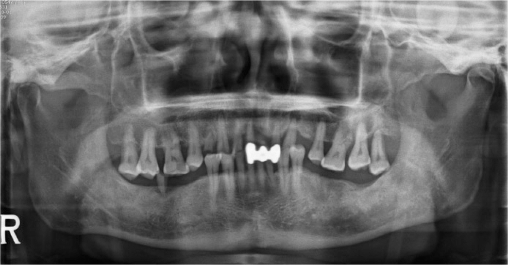
Fig. 1. Panoramic radiograph before treatment.
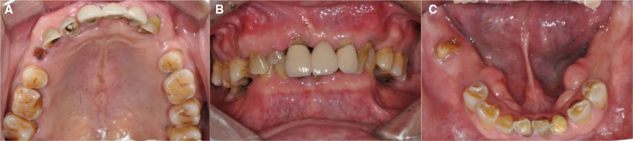
Fig. 2. Intraoral photograph before treatment. (A) Maxilla, (B) Frontal view, (C) Mandible.
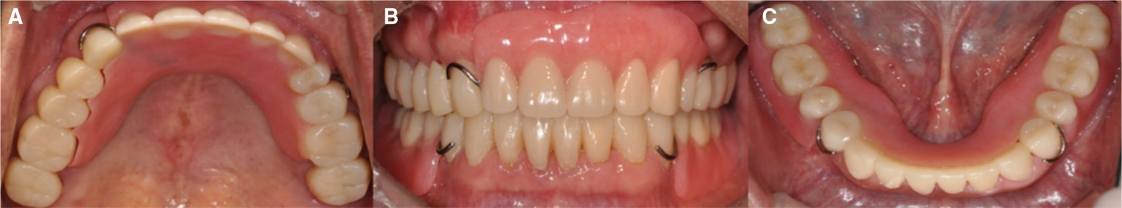
Fig. 3. Provisional restoration. (A) Maxilla, (B) Frontal view, (C) Mandible.

Fig. 4. Final restoration except for maxillary anteriors. (A) Maxilla, (B) Frontal view, (C) Mandible.

Fig. 5. T-Scan examination after posterior final restorations. (A) Occlusal contact was evenly distributed except for maxillary anterior edentulous area at MICP. The graph showed 60:40 force distribution in right (red line) and left side (green line). The occlusal time (OT) was 0.17s and disclusion time (DT) was 0.26s within the normal range, (B) Right lateral excursion movement, (C) Left lateral excursion movement.

Fig. 6. T-Scan examination after maxillary anterior provisional restoration adjustment. (A) Occlusal contact was evenly distributed at MICP and the COF was formed close to the center of the arch. The graph showed 50:50 force distribution in right (red) and left (green) side. Green and red line overlapped each other at center of graph. The occlusal time (OT) was 0.18s and disclusion time (DT) was 0.24s within the normal range, (B) Canine guidance without nonworking side contacts at right lateral excursion after adjustment. The graph showed 100:0 force distribution in right (red) and left (green) side, (C) Group function without nonworking side contacts at left lateral excursion after adjustment. The graph showed 0:100 force distribution in right (red) and left (green) side, (D) Occlusal contact was distributed on multiple teeth at the beginning of the protrusion, (E) Protrusion progressed according to the guide of the maxillary anterior teeth, (F) Occlusal contact exists only in maxillary central incisors at the end of the protrusion.
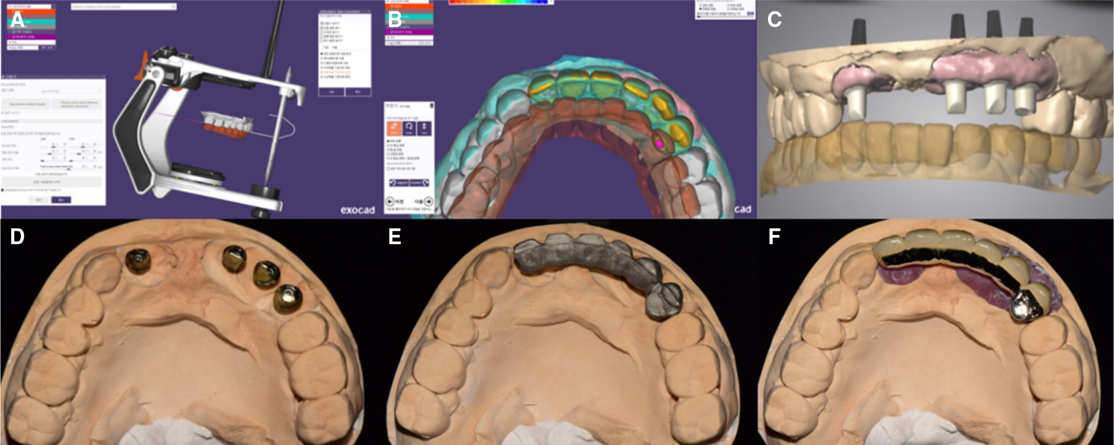
Fig. 7. Laboratory procedures using Exocad software. (A) Scanned provisional model (gray) and antagonistic teeth model (brown), (B) Superimposition and design of definitive prosthesis, (C) Design of customized abutment, (D) Fabrication of titanium customized abutment, (E) Fabrication of metal coping, (F) Definitive implant supported PFM bridge.
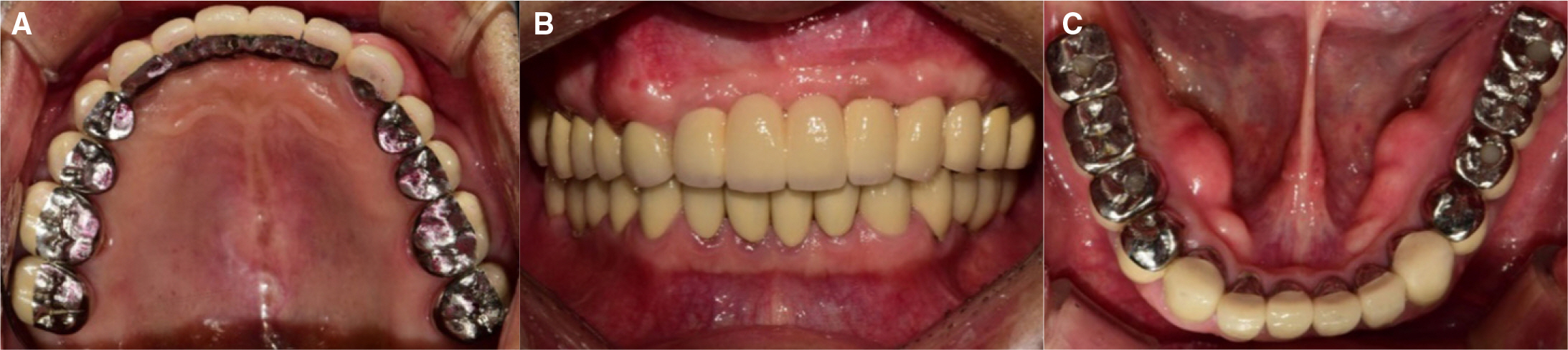
Fig. 8. Final restoration of maxillary anterior area. (A) Maxilla, (B) Frontal view, (C) Mandible.
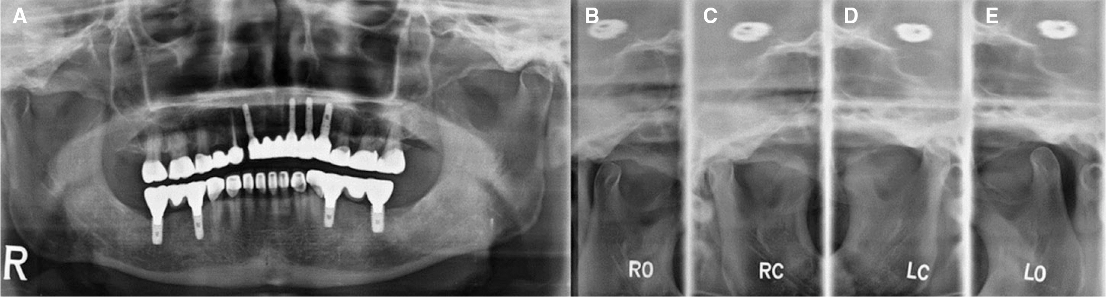
Fig. 9. Panoramic radiograph and TMJ series after final restoration. (A) Panorama (B) Rt. Opening, (C) Rt. Close, (D) Lt. Close, (E) Lt. Opening.
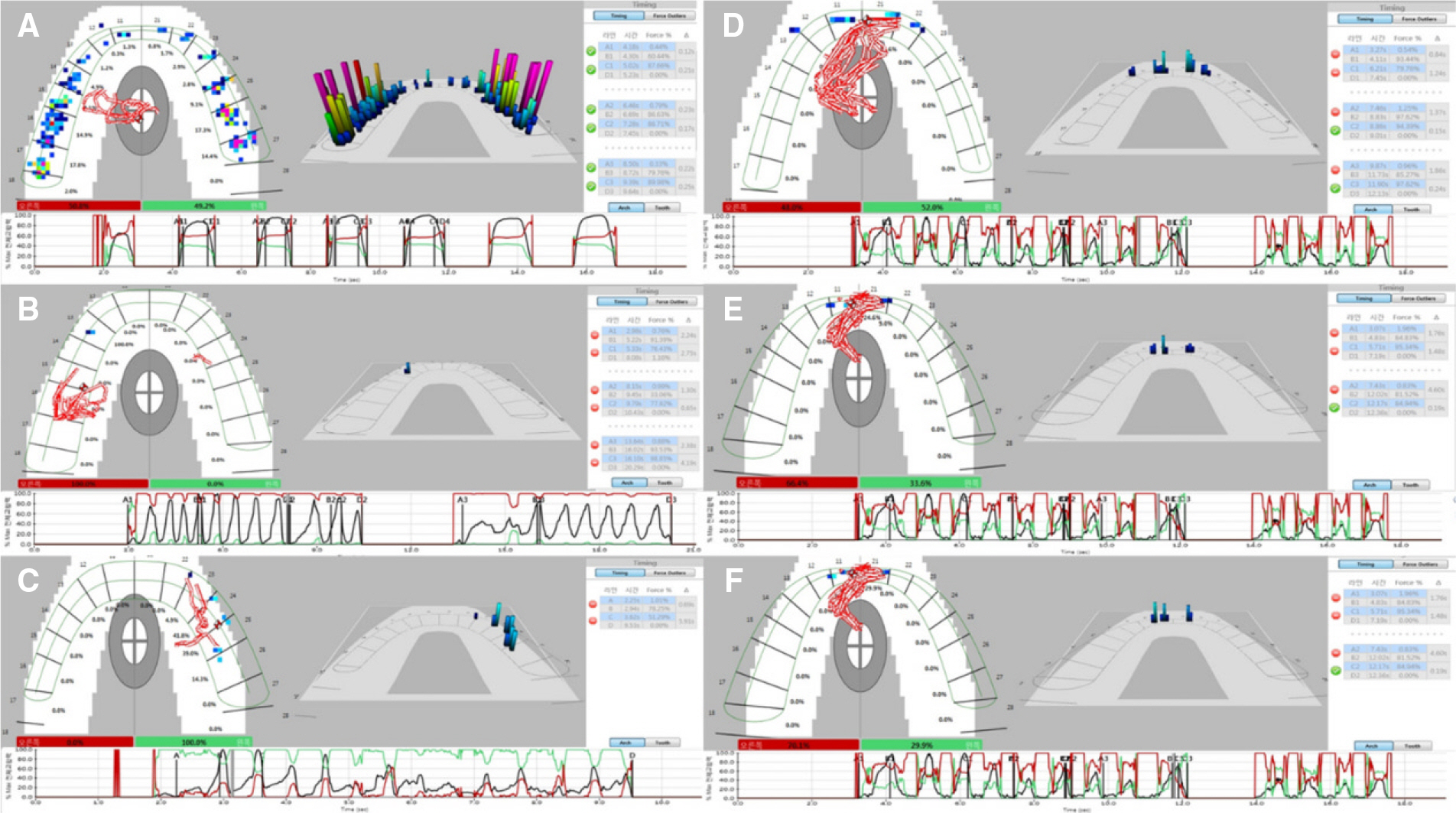
Fig. 10. T-scan examination after final restoration. (A) Occlusal contact was evenly distributed at Maximal Intercuspal Position (MICP) and the Center of Force (COF) was formed close to the center of the arch. The graph showed 50:50 force distribution in right (red) and left (green) side. The occlusal time (OT) and disclusion time (DT) values for all cycles are within the normal range (OT: 0.12s, 0.23s, 0.22s, DT: 0.21s, 0.17s, 0.25s), (B) Canine guidance without nonworking side contacts at right lateral excursion. The graph showed 100:0 force distribution in right (red) and left (green) side, (C) Group function on canine and premolars without nonworking side contacts at left lateral excursion. The graph showed 0:100 force distribution in right (red) and left (green) side, (D) Occlusal contact was evenly distributed on anterior teeth at the beginning of the protrusion, (E) Protrusion progressed according to the guide of the maxillary anterior teeth, (F) Occlusal contact exists only in maxillary central incisors at the end of the protrusion.
Reference
-
1.Carey JP., Craig M., Kerstein RB., Radke J. Determining a relationship between applied occlusal load and articulating paper mark area. Open Dent J. 2007. 1:1–7.
Article2.Kerstein RB., Grundset K. Obtaining measurable bilateral simultaneous occlusal contacts with computer-analyzed and guided occlusal adjustments. Quintessence Int. 2001. 32:7–18.3.Maness WL., Benjamin M., Podoloff R., Bobick A., Golden RF. Computerized occlusal analysis: a new technology. Quintessence Int. 1987. 18:287–92.4.Mizui M., Nabeshima F., Tosa J., Tanaka M., Kawazoe T. Quantitative analysis of occlusal balance in intercuspal position using the T-Scan system. Int J Prosthodont. 1994. 7:62–71.5.Schuyler CH. The function and importance of incisal guidance in oral rehabilitation. J Prosthet Dent. 1963. 13:1011–29.
Article6.Willis FM. Features of the face involved in full denture prosthesis. Dent Cosmos. 1935. 77:851–4.7.Nelson SJ. Wheeler's dental anatomy, physiology and occlusion. Elsevier Health Sciences;2014.8.Fayz F., Eslami A., Graser GN. Use of anterior teeth measurements in determining occlusal vertical dimension. J Prosthet Dent. 1987. 58:317–22.
Article9.Thumati P. Digital analysis of occlusion using T-Scan III in orthodontics. J Indian Orthod Soc. 2016. 50:196–201.
Article10.Becker CM., Kaiser DA., Jones JD. Guidelines for splinting implants. J Prosthet Dent. 2000. 84:210–4.
Article11.The glossary of prosthodontic terms. J Prosthet Dent. 2005. 94:10–92.12.Koos B., Godt A., Schille C., Göz G. Precision of an instrumentation-based method of analyzing occlusionand its resulting distribution of forces in the dental arch. J Orofac Orthop. 2010.
- Full Text Links
-
- Actions
-
Cited
- CITED
-
- Close
- Share
-
- Similar articles
-
- Reinforcing the retention of provisional restoration using provisional implant on maxillary anterior region: clinical case report
- Digital technique in diagnosis and restoration of maxillary anterior implant: a case report
- Esthetic restoration in continuous maxillary anterior area using immediate implant placement: A case report
- Full mouth implant-supported fixed prosthesis restoration of an edentulous maxillary patient using computer-guided implant surgery
- Full mouth rehabilitation of an edentulous patient using maxillary complete denture and mandibular implant supported fixed prostheses: a case report
