Transarterial Balloon-assisted Onyx Embolization of Intracranial Arteriovenous Malformations Using a Dual-lumen Balloon Microcatheter: Two Case Reports
- Affiliations
-
- 1Department of Neuroradiology, CHA Bundang Medical Center, CHA University School of Medicine, Seongnam, Korea.
- 2Department of Neurosurgery, CHA Bundang Medical Center, CHA University School of Medicine, Seongnam, Korea. tgkim@chamc.co.kr
- 3Department of Neurosurgery, Seoul Medical Center, Seoul, Korea.
- KMID: 2393507
- DOI: http://doi.org/10.7461/jcen.2017.19.3.223
Abstract
- The Onyx system has been well established in recent years as a very important material in the treatment of arteriovenous malformations (AVMs). When using the Onyx, it is essential to wait for the creation of a plug around the tip of the catheter, which enables the effective forward penetration of Onyx. Recent reports have shown that the introduction of a dimethyl sulfoxide compatible dual-lumen balloon microcatheter improves the efficiency of AVM embolization. We report our recent experience of two cases of intracranial AVM embolization using Onyx and the transarterial balloon-assisted technique. In both cases, the procedures were successfully performed and the nidus of the AVM was totally occluded in a relatively short time. This technique may enable immediate forward flow and penetration of Onyx without concern about reflux. It may also reduce the procedure time and increase the angiographic occlusion rate. Navigation of the dual-lumen balloon microcatheter nevertheless remains a challenge.
Keyword
MeSH Terms
Figure
-
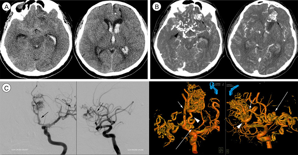
Fig. 1 A 49-year-old male patient with ruptured AVM. (A) Brain CT scan revealed diffuse SAHs in basal cisterns and large IVHs in the entire ventricles and enlarged ventricles. (B) Brain CTA showed AVM (arrows) at left lower frontal region, which was drained by enlarged veins(arrow head). (C) Conventional cerebral angiography showed AVM, which was supplied by left frontopolar artery (black and white short arrows) and the single drainage vein(white arrow heads) showed the severe ectasia, tortuosity and even varix (white long arrows). AVM = arteriovenous malformation; CT = computed tomography; CTA = computed tomography angiography.
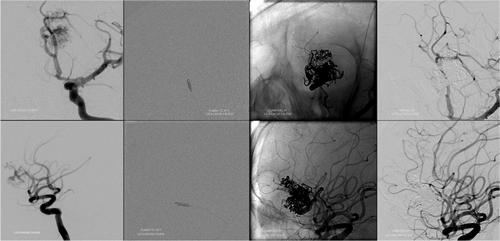
Fig. 2 Transarterial balloon-assisted Onyx embolization of intracranial AVM using dual-lumen balloon microcatheter. First column to the left: Working view anteriorposterior and lateral view, Second column: Ballooning, Third column: After completion of Onyx injection, and Fourth column: Angiography after Onyx embolization revealed the total occlusion of the AVM. AVM = arteriovenous malformation.
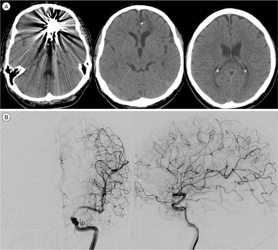
Fig. 3 Follow-up brain CT and angiograpy. (A) At two months after the hemorrhages, a brain CT scan showed no abnormal findings. (B) Seven months after the hemorrhages, the patient underwent cerebral angiography which revealed the AVM nidus remained totally occluded. CT = computed tomography; AVM = arteriovenous malformation.
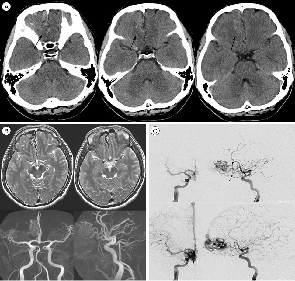
Fig. 4 A 37-year-old male patient with unruptured AVM. (A) A brain CT scan revealed mild hyperdense lesion at right medial frontal base (black arrows). (B) Brain MRI and MRA showed AVM at right frontal pole region. (C) Conventional cerebral angiography showed AVM at right frontal pole region, which was supplied by right medial oribitofrontal (black arrow) and frontopolar arteries (arrowhead) and drained by superficial cerebral vein to superior sagittal sinus. CT = computed tomography; MRI = magnetic resonance imaging, MRA = magnetic resonance angiography; AVM = arteriovenous malformation.
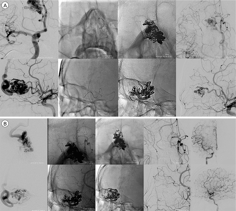
Fig. 5 Transarterial balloon-assisted Onyx embolization of intracranial AVM using dual-lumen balloon microcatheter. (A) Via the right medial orbitofrontal artery. First column to the left: Working view anteriorposterior and lateral view, Second column: After balloon catheter insertion, Third column: After completion of Onyx injection, and Fourth column: Angiography after Onyx embolization revealed the occlusion of lower two thirds of the AVM nidus. (B) Via the right frontopolar artery. First column to the left: Working view anteriorposterior and lateral view, Second column: After balloon catheter insertion, Third column: After completion of Onyx injection, Fourth column: After completion of Onyx injection, and Fifth column: Angiography after Onyx embolization revealed the total occlusion of the AVM nidus. AVM = arteriovenous malformation.
Cited by 1 articles
-
Balloon-Assisted Coil Embolization and Balloon Angioplasty for Post Subarachnoid Hemorrhage Vasospasm: Initial Experience with Scepter Mini Balloon
Ioannis Ioannidis, Antonis Adamou, Nikolaos Nasis, Marianna Vlychou, Nektarios Poullos
Neurointervention. 2022;17(2):110-114. doi: 10.5469/neuroint.2022.00171.
Reference
-
1. Abud DG, Riva R, Nakiri GS, Padovani F, Khawaldeh M, Mounayer C. Treatment of brain arteriovenous malformations by double arterial catheterization with simultaneous injection of Onyx: retrospective series of 17 patients. AJNR Am J Neuroradiol. 2011; 01. 32(1):152–158.
Article2. Andreou A, Ioannidis I, Nasis N. Transarterial balloon-assisted glue embolization of high-flow arteriovenous fistulas. Neuroradiology. 2008; 03. 50(3):267–272.
Article3. Brassel F, Solymosi L. An unusual congenital arteriovenous fistula of the vertebral artery and its embolization by a detachable balloon catheter. Neurosurg Rev. 1988; 11(1):99–101.
Article4. Chiu AH, Aw G, Wenderoth JD. Double-lumen arterial balloon catheter technique for Onyx embolization of dural arteriovenous fistulas: initial experience. J Neurointerv Surg. 2014; 06. 6(5):400–403.
Article5. Dabus G, Linfante I, Martínez-Galdámez M. Endovascular treatment of dural arteriovenous fistulas using dual lumen balloon microcatheter: technical aspects and results. Clin Neurol Neurosurg. 2014; 02. 117:22–27.
Article6. Deng JP, Zhang T, Li J, Yu J, Zhao ZW, Gao GD. Treatment of dural arteriovenous fistula by balloon-assisted transarterial embolization with Onyx. Clin Neurol Neurosurg. 2013; 10. 115(10):1992–1997.
Article7. Fifi J, Niimi Y, Berenstein A. Onyx embolization of an extensive mandibular arteriovenous malformation via a dual lumen balloon catheter: a technical case report. J Neurointerv Surg. 2013; 03. 5(2):e5.
Article8. Jagadeesan BD, Grigoryan M, Hassan AE, Grande AW, Tummala RP. Endovascular balloon-assisted embolization of intracranial and cervical arteriovenous malformations using dual-lumen coaxial balloon microcatheters and Onyx: initial experience. Neurosurgery. 2013; 12. 73:2 Suppl Operative. ons238–ons243. discussion ons243.9. Moret J, Lasjaunias P, Doyon D. Occipital approach for treatment of arteriovenous malformation of the vertebral artery by balloon occlusion. Neuroradiology. 1979; 05. 17(5):269–273.
Article10. Mounayer C, Hammami N, Piotin M, Spelle L, Benndorf G, Kessler I, et al. Nidal embolization of brain arteriovenous malformations using Onyx in 94 patients. AJNR Am J Neuroradiol. 2007; 03. 28(3):518–523.11. Newman CB, Hu YC, McDougall CG, Albuquerque FC. Balloon-assisted Onyx embolization of cerebral single-channel pial arteriovenous fistulas. J Neurosurg Pediatr. 2011; 06. 7(6):637–642.
Article12. Paramasivam S, Niimi Y, Fifi J, Berenstein A. Onyx embolization using dual-lumen balloon catheter: initial experience and technical note. J Neuroradiol. 2013; 10. 40(4):294–302.
Article13. Pierot L, Cognard C, Herbreteau D, Fransen H, van Rooij WJ, Boccardi E, et al. Endovascular treatment of brain arteriovenous malformations using a liquid embolic agent: results of a prospective, multicentre study (BRAVO). Eur Radiol. 2013; 10. 23(10):2838–2845.
Article14. Serbinenko FA. Catheterization and occlusion of major cerebral vessels and prospects for the development of vascular neurosurgery. Vopr Neirokhir. 1971; Sep-Oct. 35(5):17–27.15. Shi ZS, Loh Y, Duckwiler GR, Jahan R, Vinuela F. Balloon-assisted transarterial embolization of intracranial dural arteriovenous fistulas. J Neurosurg. 2009; 05. 110(5):921–928.
Article16. Spiotta AM, Hughes G, Masaryk TJ, Hui FK. Balloonaugmented Onyx embolization of a dural arteriovenous fistula arising from the neuromeningeal trunk of the ascending pharyngeal artery: technical report. J Neurointerv Surg. 2011; 09. 3(3):300–303.
Article17. Takeda R, Usami T, Nakagawara J, Fujiwara H, Sato S, Hyogo T, et al. Clinical application of cerebral endovascular balloon catheter for arteriovenous malformation. Case report. Neurol Med Chir (Tokyo). 1984; 03. 24(3):194–201.18. Taki W, Handa H, Yonekawa Y, Yamagata S, Miyake H, Matsuda I, et al. Detachable balloon catheter systems for embolization of cerebrovascular lesions. Neurol Med Chir (Tokyo). 1981; 07. 21(7):709–719.
Article19. Trattnig S, Samec P, Zeiler K. Balloon catheter technic and embolization--new therapeutic approaches in cerebral and spinal blood vessel malformations. Wien Klin Wochenschr. 1985; 08. 97(16):653–658.20. van Rooij WJ, Sluzewski M, Beute GN. Brain AVM embolization with Onyx. AJNR Am J Neuroradiol. 2007; 01. 28(1):172–177.21. Weber W, Kis B, Siekmann R, Kuehne D. Endovascular treatment of intracranial arteriovenous malformations with onyx: technical aspects. AJNR Am J Neuroradiol. 2007; 02. 28(2):371–377.
- Full Text Links
-
- Actions
-
Cited
- CITED
-
- Close
- Share
-
- Similar articles
-
- Retained Microcatheter after Onyx Embolization of Intracranial Arteriovenous Malformation
- Onyx Embolization of Dural Arteriovenous Fistula, using Scepter C Balloon Catheter: a Case Report
- Endovascular Treatment of Scalp Arteriovenous Fistula: Transvenous Onyx Embolization with Balloon Occlusion
- Cervical spinal extradural arteriovenous fistula successfully treated using transarterial balloon-assisted coil embolization
- Complication Associated with Onyx Embolization of Spinal Cord Arteriovenous Malformation
