Bronchial Washing Cytology of Pulmonary Langerhans Cell Histiocytosis: A Case Report
- Affiliations
-
- 1Department of Pathology, Yonsei University Wonju College of Medicine, Wonju, Korea. soonheej@yonsei.ac.kr
- KMID: 2392588
- DOI: http://doi.org/10.4132/jptm.2017.02.15
Abstract
- No abstract available.
MeSH Terms
Figure
-
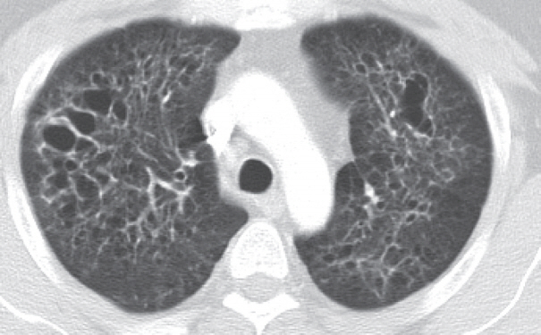
Fig. 1. Chest computed tomography findings. There are multiple, variable-sized, irregularly-shaped cysts and centrilobular nodules in both lungs.
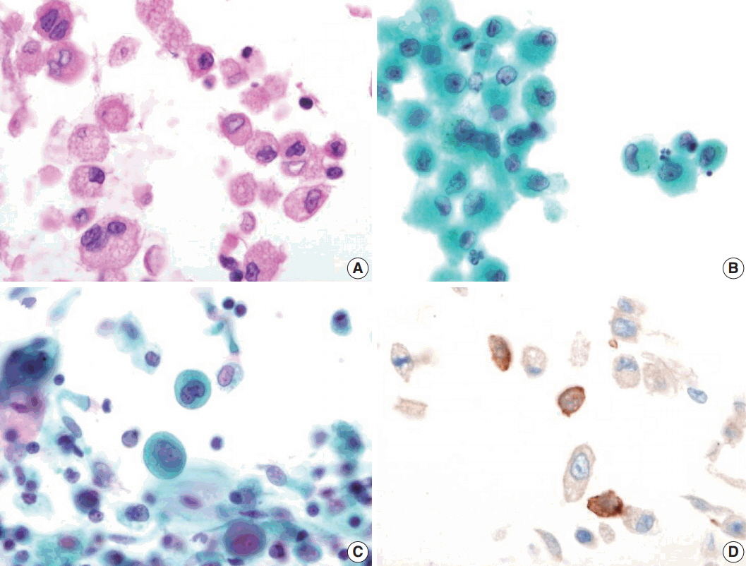
Fig. 2. Bronchial washing cytology. Bronchial washing cytology reveals many Langerhans cells. (A–C) The cells have abundant pale granular cytoplasm with a convoluted irregular nucleus and fine chromatin pattern (A, cell block; B, Thin prep; C, conventional smear). (D) Immunohistochemical staining for CD1a is positive in the Langerhans cells.
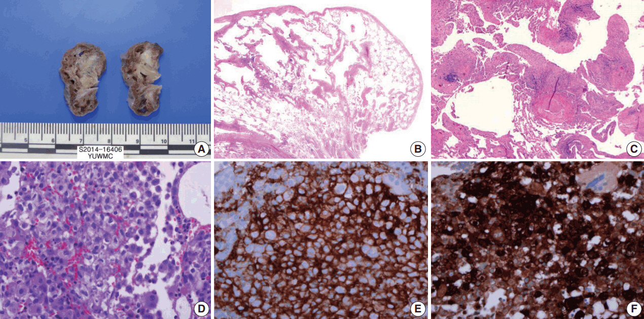
Fig. 3. Wedge resected lung via video-assisted thoracoscopic surgery. (A) The cut surface of the wedge-resected lung shows multiple numerous cystic spaces with white gray stellate fibrous scars. (B, C) There are multiple cystic spaces with diffuse thickening, cellular infiltration, and fibrous tissue. (D) The infiltrated cells have pale eosinophilic cytoplasm with grooved or infolded nuclei. (E, F) Immunohistochemical staining for CD1a and S-100 protein is positive in the proliferating cells.
Reference
-
1. Tazi A. Adult pulmonary Langerhans’ cell histiocytosis. Eur Respir J. 2006; 27:1272–85.
Article2. Yousem SA, Colby TV, Chen YY, Chen WG, Weiss LM. Pulmonary Langerhans' cell histiocytosis: molecular analysis of clonality. Am J Surg Pathol. 2001; 25:630–6.3. Takizawa Y, Taniuchi N, Ghazizadeh M, et al. Bronchoalveolar lavage fluid analysis provides diagnostic information on pulmonary Langerhans cell histiocytosis. J Nippon Med Sch. 2009; 76:84–92.
Article4. Sharma S, Dey P. Childhood pulmonary langerhans cell histiocytosis in bronchoalveolar lavage: a case report along with review of literature. Diagn Cytopathol. 2016; 44:1102–6.
Article5. Lee SR, Suh JH, Cha HJ, Kim YM, Choi HJ. Fine needle aspiration cytology of Langerhans cell histiocytosis of mandible: a case report. Korean J Pathol. 2010; 44:106–9.6. Ha SY, Kim MJ, Kim GY, Cho HY, Chung DH, Kim NR. Fine needle aspiration cytology of Langerhans cell histiocytosis in a lymph node: a case report. Korean J Cytopathol. 2007; 18:87–91.7. Kumar N, Sayed S, Vinayak S. Diagnosis of Langerhans cell histiocytosis on fine needle aspiration cytology: a case report and review of the cytology literature. Patholog Res Int. 2011; 2011:439518.
Article8. Auerswald U, Barth J, Magnussen H. Value of CD-1-positive cells in bronchoalveolar lavage fluid for the diagnosis of pulmonary histiocytosis X. Lung. 1991; 169:305–9.
Article9. Refabert L, Rambaud C, Mamou-Mani T, Scheinmann P, de Blic J. Cd1a-positive cells in bronchoalveolarlavage samples from children with Langerhans cell histiocytosis. J Pediatr. 1996; 129:913–5.10. Kilpatrick SE. Fine needle aspiration biopsy of Langerhans cell histiocytosis of bone: are ancillary studies necessary for a “definitive diagnosis”? Acta Cytol. 1998; 42:820–3.
- Full Text Links
-
- Actions
-
Cited
- CITED
-
- Close
- Share
-
- Similar articles
-
- Spontaneous Pneumothorax due to Pulmonary Invasion in Multisystemic Langerhans Cell Histiocytosis: A case report
- Pulmonary Langerhans Cell Histiocytosis Accompanied by Active Pulmonary Tuberculosis
- A Case of Pulmonary Langerhans Cell Histiocytosis with Pneumothorax
- Fine Needle Aspiration Cytology of Langerhans' Cell Histiocytosis in the Lymph Node
- Radiologic manifestation of pulmonary Langerhans' cell histiocytosis
