Imaging Sci Dent.
2017 Mar;47(1):39-44. 10.5624/isd.2017.47.1.39.
Prevalence and location of the posterior superior alveolar artery using cone-beam computed tomography
- Affiliations
-
- 1Department of Periodontology, Faculty of Dentistry, Shahed University, Tehran, Iran. ferial2002@yahoo.com
- 2Department of Oral and Maxillofacial Radiology, Faculty of Dentistry, Shahed University, Tehran, Iran.
- 3Nouri's Dental Clinic, Tehran, Iran.
- KMID: 2391405
- DOI: http://doi.org/10.5624/isd.2017.47.1.39
Abstract
- PURPOSE
Insufficient knowledge of the anatomy of the maxillary sinuses prior to sinus graft surgery may lead to perioperative or postoperative complications. This study sought to characterize the position of the posterior superior alveolar artery (PSAA) within the maxillary sinuses using cone-beam computed tomography (CBCT).
MATERIALS AND METHODS
A total of 300 patients with edentulous posterior maxillae, including 138 females and 162 males with an age range of 33-86 years, who presented to a radiology clinic between 2013 and 2015 were enrolled in this retrospective cross-sectional study. The distance from the inferior border of the PSAA to the alveolar crest according to the residual ridge classification by Lekholm and Zarb, the distance from the PSAA to the nasal septum and zygomatic arch, and the diameter and position of the PSAA were all assessed on patients' CBCT scans. The data were analyzed using the Mann-Whitney test and the t-test.
RESULTS
The PSAA was detected on the CBCT scans of 87% of the patients; it was located beneath the sinus membrane in 47% of cases and was intraosseous in 47% of cases. The diameter of the artery was between 1 and 2 mm in most patients (72%). The mean diameter of the artery was 1.29±0.39 mm, and the mean distances from the PSAA to the zygomatic arch, nasal septum, and alveolar crest were 22.59±4.89 mm, 26.51±3.52 mm, and 16.7±3.96 mm, respectively.
CONCLUSION
The likelihood of detecting the PSAA on CBCT scans is high; its location is intraosseous or beneath the sinus membrane in most patients. Determining the exact location of the PSAA on CBCT scans preoperatively can help prevent it from being damaged during surgery.
MeSH Terms
Figure
-
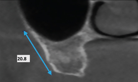
Fig. 1 Distance from the lower border of the artery to the alveolar crest.
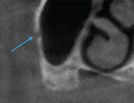
Fig. 2 Coronal view of the maxillary sinus, showing the intraosseous artery.

Fig. 3 Coronal view of the maxillary sinus, showing the artery (arrow), which is below the membrane.
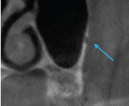
Fig. 4 Coronal view of the maxillary sinus, showing the artery (arrow), which runs over the external cortex of the lateral sinus wall.

Fig. 5 Distance from the artery to the zygomatic arch (A) and to the nasal septum (B).
Reference
-
1. Newman MG, Takei HH, Klokkevold PR, Carranza FA. Carranza's clinical periodontology. 12th ed. St. Louis, MO: Saunders;2015.2. Boyne PJ, James RA. Grafting of the maxillary sinus floor with autogenous marrow and bone. J Oral Surg. 1980; 38:613–616.3. Pjetursson BE, Tan WC, Zwahlen M, Lang NP. A systematic review of the success of sinus floor elevation and survival of implants inserted in combination with sinus floor elevation. J Clin Periodontol. 2008; 35:8 Suppl. 216–240.
Article4. Solar P, Geyerhofer U, Traxler H, Windisch A, Ulm C, Watzek G. Blood supply to the maxillary sinus relevant to sinus floor elevation procedures. Clin Oral Implants Res. 1999; 10:34–44.
Article5. Ella B, Sedarat C, Noble Rda C, Normand E, Lauverjat Y, Siberchicot F, et al. Vascular connections of the lateral wall of the sinus: surgical effect in sinus augmentation. Int J Oral Maxillofac Implants. 2008; 23:1047–1052.6. Ilgüy D, Ilgüy M, Dolekoglu S, Fisekcioglu E. Evaluation of the posterior superior alveolar artery and the maxillary sinus with CBCT. Braz Oral Res. 2013; 27:431–437.7. Arai Y, Tammisalo E, Iwai K, Hashimoto K, Shinoda K. Development of a compact computed tomographic apparatus for dental use. Dentomaxillofac Radiol. 1999; 28:245–248.
Article8. Mozzo P, Procacci C, Tacconi A, Martini PT, Andreis IA. A new volumetric CT machine for dental imaging based on the cone-beam technique: preliminary results. Eur Radiol. 1998; 8:1558–1564.
Article9. Ludlow JB, Ivanovic M. Comparative dosimetry of dental CBCT devices and 64-slice CT for oral and maxillofacial radiology. Oral Surg Oral Med Oral Pathol Oral Radiol Endod. 2008; 106:106–114.
Article10. Lekholm U, Zarb GA. Patient selection and preparation. In : Brånemark PI, George AZ, Albrektsson T, editors. Tissue-integrated prostheses: osseointegration in clinical dentistry. Chicago, IL: Quintessence;1985. p. 199–209.11. Elian N, Wallace S, Cho SC, Jalbout ZN, Froum S. Distribution of the maxillary artery as it relates to sinus floor augmentation. Int J Oral Maxillofac Implants. 2005; 20:784–787.12. Mardinger O, Abba M, Hirshberg A, Schwartz-Arad D. Prevalence, diameter and course of the maxillary intraosseous vascular canal with relation to sinus augmentation procedure: a radiographic study. Int J Oral Maxillofac Surg. 2007; 36:735–738.
Article13. Rosano G, Taschieri S, Gaudy JF, Del Fabbro M. Maxillary sinus vascularization: a cadaveric study. J Craniofac Surg. 2009; 20:940–943.14. Güncü GN, Yildirim YD, Wang HL, Tözüm TF. Location of posterior superior alveolar artery and evaluation of maxillary sinus anatomy with computerized tomography: a clinical study. Clin Oral Implants Res. 2011; 22:1164–1167.
Article15. Kim JH, Ryu JS, Kim KD, Hwang SH, Moon HS. A radiographic study of the posterior superior alveolar artery. Implant Dent. 2011; 20:306–310.
Article16. Kim MJ, Jung UW, Kim CS, Kim KD, Choi SH, Kim CK, et al. Maxillary sinus septa: prevalence, height, location, and morphology. A reformatted computed tomography scan analysis. J Periodontol. 2006; 77:903–908.
Article17. Monje A, Catena A, Monje F, Gonzalez-Garcia R, Galindo-Moreno P, Suarez F, et al. Maxillary sinus lateral wall thickness and morphologic patterns in the atrophic posterior maxilla. J Periodontol. 2014; 85:676–682.
Article
- Full Text Links
-
- Actions
-
Cited
- CITED
-
- Close
- Share
-
- Similar articles
-
- Anatomic evaluation of the posterior superior alveolar artery using cone-beam computed tomography: A systematic review and meta-analysis
- Evaluation of the posterior superior alveolar artery canal by cone-beam computed tomography in a sample of the Egyptian population
- Radiomorphometric analysis of edentulous posterior mandibular ridges in the first molar region: a cone-beam computed tomography study
- Comparative evaluation of computed tomography for dental implants on the mandibular edentulous area
- Management of root canal perforation by using cone-beam computed tomography
