Imaging Sci Dent.
2017 Mar;47(1):33-37. 10.5624/isd.2017.47.1.33.
Cone-beam computed tomography analysis of accessory maxillary ostium and Haller cells: Prevalence and clinical significance
- Affiliations
-
- 1Department of Oral Medicine and Radiology, Nair Hospital Dental College, Mumbai, Maharashtra, India. ibrahimali2589@gmail.com
- 2Department of Conservative Dentistry and Endodontics, Nair Hospital Dental College, Mumbai, Maharashtra, India.
- KMID: 2391404
- DOI: http://doi.org/10.5624/isd.2017.47.1.33
Abstract
- PURPOSE
This study aimed to evaluate the prevalence of Haller cells and accessory maxillary ostium (AMO) in cone-beam computed tomography (CBCT) images, and to analyze the relationships among Haller cells, AMO, and maxillary sinusitis.
MATERIALS AND METHODS
Volumetric CBCT scans from 201 patients were retrieved from our institution's Digital Imaging and Communications in Medicine archive folder. Two observers evaluated the presence of Haller cells, AMO, and maxillary sinusitis in the CBCT scans.
RESULTS
AMO was observed in 114 patients, of whom 27 (23.7%) had AMO exclusively on the right side, 26 (22.8%) only on the left side, and 61 (53.5%) bilaterally. Haller cells were identified in 73 (36.3%) patients. In 24 (32.9%) they were present exclusively on the right side, in 17 (23.3%) they were only present on the left side, and in 32 (43.8%) they were located bilaterally. Of the 73 (36.3%) patients with Haller cells, maxillary sinusitis was also present in 50 (68.5%). On using chi-square test, a significant association was observed between AMO and maxillary sinusitis in the presence of Haller cells.
CONCLUSION
Our results showed AMO and Haller cells to be associated with maxillary sinusitis. This study provides evidence for the usefulness of CBCT in imaging the bony anatomy of the sinonasal complex with significantly higher precision and a smaller radiation dose.
MeSH Terms
Figure
-
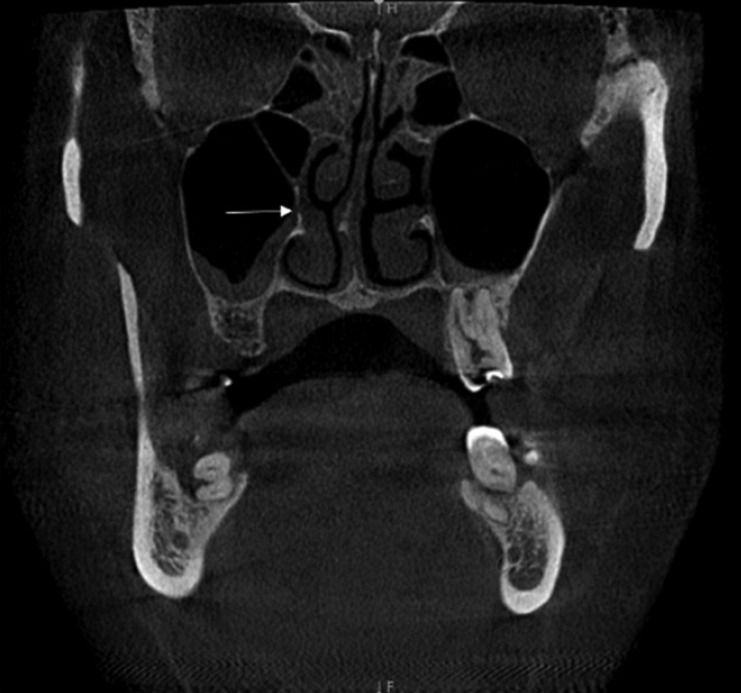
Fig. 1 A coronal cone-beam computed tomography image demonstrates accessory maxillary ostium in the right maxillary sinus with ipsilateral maxillary sinusitis (white arrow).
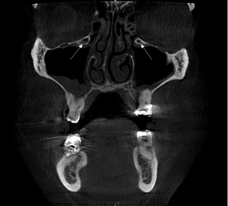
Fig. 2 A coronal cone-beam computed tomography image shows the bilateral presence of Haller cells with maxillary sinusitis (white arrow).
Reference
-
2. Lloyd GA. CT of the paranasal sinuses: study of a control series in relation to endoscopic sinus surgery. J Laryngol Otol. 1990; 104:477–481. PMID: 2376707.
Article3. Yenigun A, Fazliogullari Z, Gun C, Uysal II, Nayman A, Karabulut AK. The effect of the presence of the accessory maxillary ostium on the maxillary sinus. Eur Arch Otorhinolaryngol. 2016; 273:4315–4319. PMID: 27300297.
Article4. Genc S, Ozcan M, Titiz A, Unal A. Development of maxillary accessory ostium following sinusitis in rabbits. Rhinology. 2008; 46:121–124. PMID: 18575013.5. Mladina R, Vuković K, Poje G. The two holes syndrome. Am J Rhinol Allergy. 2009; 23:602–604. PMID: 19958610.
Article6. Kane KJ. Recirculation of mucus as a cause of persistent sinusitis. Am J Rhinol. 1997; 11:361–369. PMID: 9768318.
Article7. Gutman M, Houser S. Iatrogenic maxillary sinus recirculation and beyond. Ear Nose Throat J. 2003; 82:61–63. PMID: 12610908.
Article8. Mathew R, Omami G, Hand A, Fellows D, Lurie A. Cone beam CT analysis of Haller cells: prevalence and clinical significance. Dentomaxillofac Radiol. 2013; 42:20130055. PMID: 23975112.
Article9. Stammberger H, Wolf G. Headaches and sinus disease: the endoscopic approach. Ann Otol Rhinol Laryngol Suppl. 1988; 134:3–23. PMID: 3140703.
Article10. Stackpole SA, Edelstein DR. The anatomic relevance of the Haller cell in sinusitis. Am J Rhinol. 1997; 11:219–223. PMID: 9209594.
Article11. Kantarci M, Karasen RM, Alper F, Onbas O, Okur A, Karaman A. Remarkable anatomic variations in paranasal sinus region and their clinical importance. Eur J Radiol. 2004; 50:296–302. PMID: 15145491.
Article12. Rafferty MA, Siewerdsen JH, Chan Y, Moseley DJ, Daly MJ, Jaffray DA, et al. Investigation of C-arm cone-beam CT-guided surgery of the frontal recess. Laryngoscope. 2005; 115:2138–2143. PMID: 16369157.
Article13. Jackman AH, Palmer JN, Chiu AG, Kennedy DW. Use of intraoperative CT scanning in endoscopic sinus surgery: a preliminary report. Am J Rhinol. 2008; 22:170–174. PMID: 18416975.
Article14. Batra PS, Kanowitz SJ, Citardi MJ. Clinical utility of intraoperative volume computed tomography scanner for endoscopic sinonasal and skull base procedures. Am J Rhinol. 2008; 22:511–515. PMID: 18954511.
Article15. Kumar H, Choudhry R, Kakar S. Accessory maxillary ostia: topography and clinical application. J Anat Soc India. 2001; 50:3–5.16. Jog M, McGarry GW. How frequent are accessory sinus ostia? J Laryngol Otol. 2003; 117:270–272. PMID: 12816215.
Article17. Earwaker J. Anatomic variants in sinonasal CT. Radiographics. 1993; 13:381–415. PMID: 8460226.
Article18. May M, Sobol SM, Korzec K. The location of the maxillary os and its importance to the endoscopic sinus surgeon. Laryngoscope. 1990; 100:1037–1042. PMID: 2215032.
Article19. Bolger WE, Butzin CA, Parsons DS. Paranasal sinus bony anatomic variations and mucosal abnormalities: CT analysis for endoscopic sinus surgery. Laryngoscope. 1991; 101:56–64. PMID: 1984551.20. Pérez-Piñas I, Sabaté J, Carmona A, Catalina-Herrera CJ, Jiménez-Castellanos J. Anatomical variations in the human paranasal sinus region studied by CT. J Anat. 2000; 197:221–227. PMID: 11005714.21. Rysz M, Bakoń L. Maxillary sinus anatomy variation and nasal cavity width: structural computed tomography imaging. Folia Morphol (Warsz). 2009; 68:260–264. PMID: 19950077.
- Full Text Links
-
- Actions
-
Cited
- CITED
-
- Close
- Share
-
- Similar articles
-
- Detection of maxillary second molar with two palatal roots using cone beam computed tomography: a case report
- Evaluation of Maxillary Sinus Using Cone-beam Computed Tomography in Patients Who Underwent Le Fort I Osteotomy
- Comparison of panoramic radiography and cone beam computed tomography for assessing the relationship between the maxillary sinus floor and maxillary molars
- A rare case of dilated invaginated odontome with talon cusp in a permanent maxillary central incisor diagnosed by cone beam computed tomography
- Prevalence of incidental paranasal sinus opacification in dental paediatric patients
