Hepatocellular Carcinoma with Vascular Central Scar
- Affiliations
-
- 1Department of Radiology, Haeundae Paik Hospital, Inje University College of Medicine, Busan, Korea. chosai81@gmail.com
- 2Department of Pathology, Haeundae Paik Hospital, Inje University College of Medicine, Busan, Korea.
- 3Department of Surgery, Haeundae Paik Hospital, Inje University College of Medicine, Busan, Korea.
- KMID: 2390794
- DOI: http://doi.org/10.4166/kjg.2017.70.3.150
Abstract
- No abstract available.
MeSH Terms
Figure
-

Fig. 1 Liver dynamic computed tomography (CT). CT images show an enhancing nodule in segment IV. The nodule shows low attenuation in precontrast image (A), enhancement in arterial phase (B) and wash out in portal and delayed phase (C, D). The central scar shows low attenuation in arterial phase and enhancement in delayed phase.
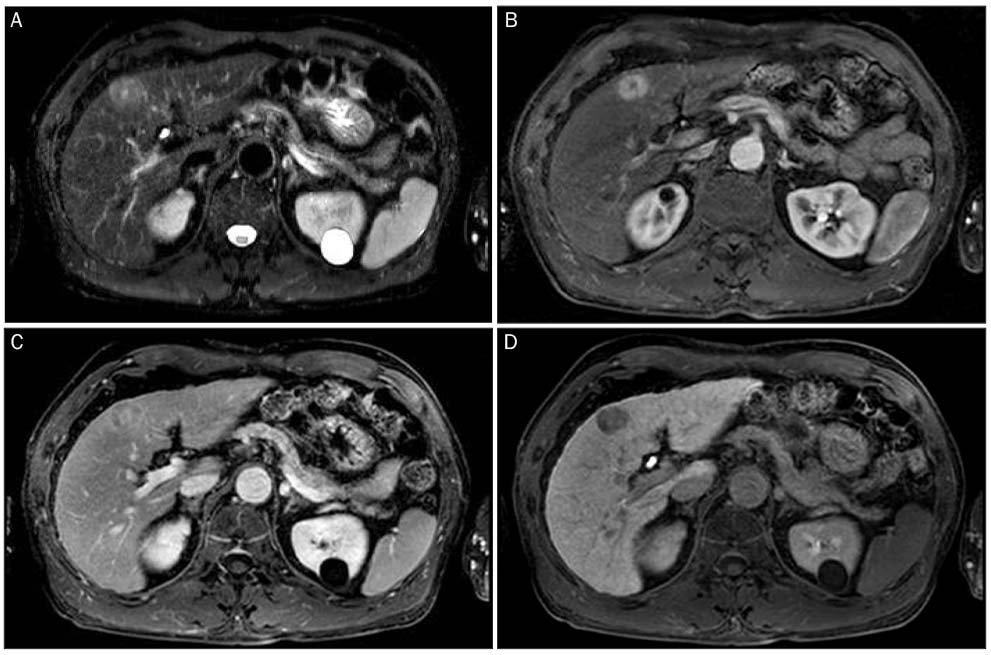
Fig. 2 Liver magnetic resonance images (MRI). (A) MRIs show a nodule with central scar showing high signal intensity on T2 weighted image. (B, C) The nodule shows scalloped margin and homogenous enhancement on arterial phase (B), low signal intensity on hepatic phase (C). The central scar shows low signal intensity on arterial phase and delayed enhancement on hepatic phase. (D) On hepatobiliary phase, the nodule shows low signal intensity.
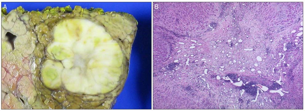
Fig. 3 Pathologic findings of surgical specimen. (A) Gross appearance of the resected liver shows a well-defined, whitish-yellow mass with a central scar. (B) Microscopic finding of the tumor shows a central scar consisting of many thin-walled blood vessels within a fibrous stroma (H&E, ×40).
Reference
-
1. Kim T, Hori M, Onishi H. Liver masses with central or eccentric scar. Semin Ultrasound CT MR. 2009; 30:418–425.2. Rummeny E, Weissleder R, Sironi S, et al. Central scars in primary liver tumors: MR features, specificity, and pathologic correlation. Radiology. 1989; 171:323–326.3. Blachar A, Federle MP, Ferris JV, et al. Radiologists’ performance in the diagnosis of liver tumors with central scars by using specific CT criteria. Radiology. 2002; 223:532–539.4. McLarney JK, Rucker PT, Bender GN, Goodman ZD, Kashitani N, Ros PR. Fibrolamellar carcinoma of the liver: radiologic-pathologic correlation. Radiographics. 1999; 19:453–471.5. Ichikawa T, Federle MP, Grazioli L, Madariaga J, Nalesnik M, Marsh W. Fibrolamellar hepatocellular carcinoma: imaging and pathologic findings in 31 recent cases. Radiology. 1999; 213:352–361.6. Hussain SM, Terkivatan T, Zondervan PE, et al. Focal nodular hyperplasia: findings at state-of-the-art MR imaging, US, CT, and pathologic analysis. Radiographics. 2004; 24:3–17. discussion 18-19.7. Yamamoto M, Ariizumi S, Yoshitoshi K, Saito A, Nakano M, Takasaki K. Hepatocellular carcinoma with a central scar and a scalloped tumor margin resembling focal nodular hyperplasia in macroscopic appearance. J Surg Oncol. 2006; 94:587–591.8. Yamauchi M, Asayama Y, Yoshimitsu K, et al. Hepatocellular carcinoma with a prominent vascular scar in the center: MR imaging findings. Radiat Med. 2006; 24:467–470.9. Quaglia A, Tibballs J, Grasso A, et al. Focal nodular hyperplasia-like areas in cirrhosis. Histopathology. 2003; 42:14–21.10. Kamel IR, Liapi E, Fishman EK. Focal nodular hyperplasia: lesion evaluation using 16-MDCT and 3D CT angiography. AJR Am J Roentgenol. 2006; 186:1587–1596.11. Kobayashi S, Matsui O, Kamura T, et al. Imaging of benign hypervascular hypatocellular nodules in alcoholic liver cirrhosis: differentiation from hypervascular hepatocellular carcinomas. J Comput Assist Tomogr. 2007; 31:557–563.12. Yoneda N, Matsui O, Kitao A, et al. Hepatocyte transporter expression in FNH and FNH-like nodule: correlation with signal intensity on gadoxetic acid enhanced magnetic resonance images. Jpn J Radiol. 2012; 30:499–508.13. Fujiwara H, Sekine S, Onaya H, Shimada K, Mikata R, Arai Y. Ring-like enhancement of focal nodular hyperplasia with hepatobiliary-phase Gd-EOB-DTPA-enhanced magnetic resonance imaging: radiological-pathological correlation. Jpn J Radiol. 2011; 29:739–743.14. Grazioli L, Bondioni MP, Heradome H, et al. Hepatocellular adenoma and focal nodular hyperplasia: value of gadoxetic acid-enhanced MR imaging in differential diagnosis. Radiology. 2012; 262:520–529.
- Full Text Links
-
- Actions
-
Cited
- CITED
-
- Close
- Share
-
- Similar articles
-
- Primary Hepatic Carcinoid Tumor: A case report
- Metastatic Omental Hepatocellular Carcinoma: Two Cases Report
- Expression of Vascular Endothelial Growth Factor (VEGF) and Microvessel Density in Hepatocellular Carcinoma
- Hepatocellular Carcinoma Arising in Hepatocellular Adenoma
- A Case of Cutaneous Acrometastasis of Hepatocellular Carcinoma to the Finger
