Is tricuspid valve really tricuspid?
- Affiliations
-
- 1Department of Anatomy, All India Institute of Medical Sciences, Bhopal, India. sunita.anatomy@aiimsbhopal.edu.in
- 2Department of Anatomy, People's College of Medical Sciences and Research Center, Bhopal, India.
- 3Department of Anatomy, Andaman & Nicobar Islands Institute of Medical Sciences, Port Blair, India.
- KMID: 2390419
- DOI: http://doi.org/10.5115/acb.2017.50.1.1
Abstract
- Advancement in imaging techniques and interventional cardiology procedures have generated renewed interest in anatomy of tricuspid valve complex. The purpose of the present study was to characterize the morphology of tricuspid valve leaflets using objective criteria. Thirty-six embalmed cadaveric hearts were utilized for the present study. Leaflet morphology was studied using newly defined criteria. Commissural zones were identified and leaflets were delineated. Presence of scallops was also recorded. Single leaflet was observed in six cases, double in 26 cases, and triple in four cases. The anterior leaflet is large with multiple scallops and frequently accrues portion of inferior leaflet. The septal leaflet is in the form of a plateau and also frequently accrues parts of inferior leaflet. The inferior leaflet rarely occurs as independent leaflet. A wide un-indented basal zone exists across the valve leaflets. The study found that the tricuspid valve is rarely tricuspid. It also generated the hypotheses that the tricuspid valve does not open completely due to presence of a wide basal zone and the valve does not close completely owing to incongruence and lack of coaptation of leaflets. The findings provide clear understanding of leaflet morphology of tricuspid valve. This will help imaging specialists for interpretation of images and cardiologists for interventional procedures. The findings also enhance our understanding of pathophysiology of conditions like functional tricuspid regurgitation.
Keyword
MeSH Terms
Figure
-
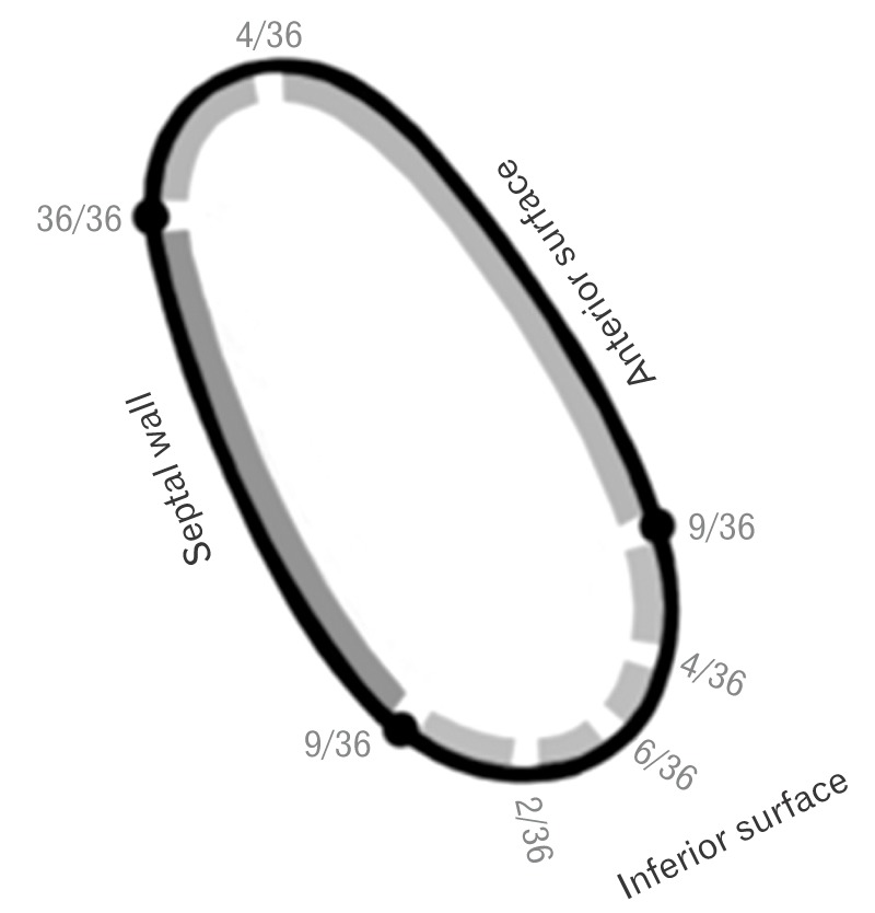
Fig. 1 Schematic diagram showing position of major commissural zone along with their frequency along the annular circumference. Gaps represent the site of commissural zone and thickened continuous areas denote the leaflets. Black dots represent conventional positions (at the junction of different surfaces of the ventricular wall) of commissures.

Fig. 2 Supraventricular leaflet (SVL) overlying the supraventricular crest between the two major commissures (asterisks).
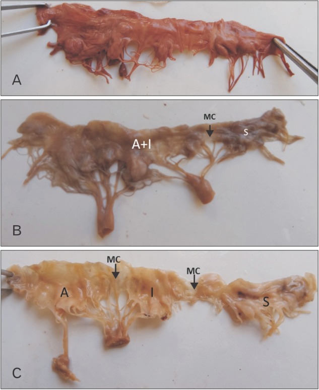
Fig. 3 Single (with wide unindented basal zone) (A), double (B), and three (C) leaflets. The valve has been cut open at the constant major commissure. MC, major commissure; A, anterior leaflet; I, inferior leaflet; S, septal leaflet; A+I, anterior has completely accrued the inferior leaflet.
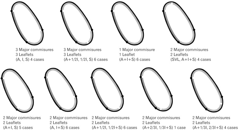
Fig. 4 Schematic diagram depicting the pattern, characterization and number of leaflets and major commissures in the hearts studied. Gaps represent the site of major commissural zone and thickened continuous areas denote the leaflets. A, anterior leaflet; I, inferior leaflet; S, septal leaflet; SVL, supraventricular leaflet.
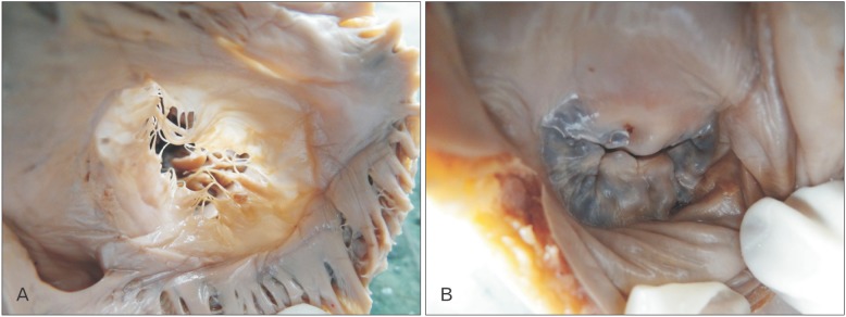
Fig. 5 Difference in coaptation of leaflets between tricuspid and mitral valve. (A) Poorly congruent tricuspid with minimum coaptation. (B) Perfectly congruent and coapted mitral valve.
Cited by 1 articles
-
Histological assessment of the human heart valves and its relationship with age
Treerat Gumpangseth, Suree Lekawanvijit, Pasuk Mahakkanukrauh
Anat Cell Biol. 2020;53(3):261-271. doi: 10.5115/acb.20.093.
Reference
-
1. Hollinshead WH. Anatomy for surgeons. Vol. 2. The thorax, abdomen and pelvis. 2nd ed. New York: Harper and Row;1971. p. 130.2. Standring S. Gray's anatomy: the anatomical basis of clinical practice. 39th ed. London: Churchill Livingstone;2005. p. 1163–1164.3. Sinnatamby CS. Last's anatomy: regional and applied. 10th ed. London: Churchill Livingstone;2001. p. 194.4. Sutton JP 3rd, Ho SY, Vogel M, Anderson RH. Is the morphologically right atrioventricular valve tricuspid? J Heart Valve Dis. 1995; 4:571–575. PMID: 8611971.5. Victor S, Nayak VM. The tricuspid valve is bicuspid. J Heart Valve Dis. 1994; 3:27–36. PMID: 8162211.6. Joudinaud TM, Flecher EM, Duran CM. Functional terminology for the tricuspid valve. J Heart Valve Dis. 2006; 15:382–388. PMID: 16784076.7. Skwarek M, Hreczecha J, Dudziak M, Jerzemowski J, Wilk B, Grzybiak M. The morphometry of the accessory leaflets of the tricuspid valve in a four cuspidal model. Folia Morphol (Warsz). 2007; 66:323–327. PMID: 18058755.8. Lama P, Tamang BK, Kulkarni J. Morphometry and aberrant morphology of the adult human tricuspid valve leaflets. Anat Sci Int. 2016; 91:143–150. PMID: 25677415.9. Rusted IE, Scheifley CH, Edwards JE, Kirklin JW. Guides to the commissures in operations upon the mitral valve. Proc Staff Meet Mayo Clin. 1951; 26:297–305. PMID: 14864669.10. Ranganathan N, Lam JH, Wigle ED, Silver MD. Morphology of the human mitral valve. II. The value leaflets. Circulation. 1970; 41:459–467. PMID: 5415983.11. Silver MD, Lam JH, Ranganathan N, Wigle ED. Morphology of the human tricuspid valve. Circulation. 1971; 43:333–348. PMID: 5544987.12. Klein AL, Burstow DJ, Tajik AJ, Zachariah PK, Taliercio CP, Taylor CL, Bailey KR, Seward JB. Age-related prevalence of valvular regurgitation in normal subjects: a comprehensive color flow examination of 118 volunteers. J Am Soc Echocardiogr. 1990; 3:54–63. PMID: 2310593.13. Rogers JH, Bolling SF. The tricuspid valve: current perspective and evolving management of tricuspid regurgitation. Circulation. 2009; 119:2718–2725. PMID: 19470900.14. Singh JP, Evans JC, Levy D, Larson MG, Freed LA, Fuller DL, Lehman B, Benjamin EJ. Prevalence and clinical determinants of mitral, tricuspid, and aortic regurgitation (the Framingham Heart Study). Am J Cardiol. 1999; 83:897–902. PMID: 10190406.15. Fukuda S, Saracino G, Matsumura Y, Daimon M, Tran H, Greenberg NL, Hozumi T, Yoshikawa J, Thomas JD, Shiota T. Three-dimensional geometry of the tricuspid annulus in healthy subjects and in patients with functional tricuspid regurgitation: a real-time, 3-dimensional echocardiographic study. Circulation. 2006; 114(1 Suppl):I492–I498. PMID: 16820625.
- Full Text Links
-
- Actions
-
Cited
- CITED
-
- Close
- Share
-
- Similar articles
-
- The Morphologic Study of the Tricuspid Valve Complex in Korean Adult Hearts
- Tricuspid Valve Re-Repair in Ebstein Anomaly Using the Cone Technique
- Permanent Pacemaker Lead Induced Severe Tricuspid Regurgitation in Patient Undergoing Multiple Valve Surgery
- Quadrangular Resection of the Tricuspid Valve
- Three-dimensional Reconstruction of Tricuspid Annulus with the Duran Ring
