Korean J Ophthalmol.
2017 Oct;31(5):460-461. 10.3341/kjo.2017.0032.
A Case of Rothia mucilaginosa Keratitis in South Korea
- Affiliations
-
- 1Department of Ophthalmology, Chonbuk National University Medical School, Jeonju, Korea. you2ic@daum.net
- 2Research Institute of Clinical Medicine, Chonbuk National University, Jeonju, Korea.
- 3Biomedical Research Institute, Chonbuk National University Hospital, Jeonju, Korea.
- KMID: 2390290
- DOI: http://doi.org/10.3341/kjo.2017.0032
Abstract
- No abstract available.
MeSH Terms
Figure
-
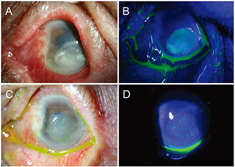
Fig. 1 Slit-lamp photographs of Rothia mucilaginosa keratitis at initial presentation. Anterior segment photograph shows severe ciliary injection and chemosis, about a 5.0 × 4.5 mm sized central corneal epithelial defect and stromal infiltration, Descemet's membrane folding, and hypopyon (A,B). Slit-lamp photographs 2 months after initial treatment. Anterior segment photographs show healed epithelial defect, fainted corneal opacity, and peripheral neovascularization (C,D).
Reference
-
1. Ramanan P, Barreto JN, Osmon DR, Tosh PK. Rothia bacteremia: a 10-year experience at Mayo Clinic, Rochester, Minnesota. J Clin Microbiol. 2014; 52:3184–3189.2. Tan R, White V, Servais G, Bryce EA. Postoperative endophthalmitis caused by Stomatococcus mucilaginosus. Clin Infect Dis. 1994; 18:492–493.3. Mattern RM, Ding J. Keratitis with Kocuria palustris and Rothia mucilaginosa in vitamin A deficiency. Case Rep Ophthalmol. 2014; 5:72–77.4. Alvarez-Ramos P, Del Moral-Ariza A, Alonso-Maroto JM, et al. First report of acute postoperative endophthalmitis caused by Rothia mucilaginosa after phacoemulsification. Infect Dis Rep. 2016; 8:6320.5. Oie S, Mochizuki K, Ishida K, et al. Case of late-onset bleb associated endophthalmitis caused by Rothia mucilaginosa. J Infect Chemother. 2016; 22:645–647.
- Full Text Links
-
- Actions
-
Cited
- CITED
-
- Close
- Share
-
- Similar articles
-
- A Case of Prosthetic Valve Endocarditis with Cerebral Hemorrhage Caused by Rothia mucilaginosa
- A case of peritoneal dialysis-associated peritonitis by Rothia mucilaginosa
- Rothia mucilaginosa Pneumonia Diagnosed by Quantitative Cultures and Intracellular Organisms of Bronchoalveolar Lavage in a Lymphoma Patient
- Molecular Identification of Clinical Rothia Isolates from Human Patients: Proposal of a Novel Rothia Species, Rothia arfidiae sp. nov.
- The Effect of Subconjunctival Injection of Tathion on Some Keratitis
