Imaging Sci Dent.
2017 Sep;47(3):175-180. 10.5624/isd.2017.47.3.175.
Prevalence of pineal gland calcification as an incidental finding in patients referred for implant dental therapy
- Affiliations
-
- 1Section of Oral and Maxillofacial Radiology, School of Dental Medicine, University of Connecticut Health Center, Farmington, CT, USA. tadinada@uchc.edu
- KMID: 2390082
- DOI: http://doi.org/10.5624/isd.2017.47.3.175
Abstract
- PURPOSE
Pineal gland calcification has been proposed to play a role in the pathogenesis of Alzheimer disease. This study evaluated the prevalence and extent of pineal gland calcification in cone-beam computed tomography (CBCT) scans of patients referred for dental implant therapy who could possibly be a vulnerable group for this condition.
MATERIALS AND METHODS
A retrospective evaluation of 500 CBCT scans was conducted. Scans that showed the area where the pineal gland was located were included. The scans were initially screened by a single observer to record the prevalence and extent of calcification. Six weeks following the completion of the study, another investigator randomly reviewed and selected 50 scans to investigate inter-observer variation, which was evaluated using reliability analysis statistics. The prevalence and measurements of the calcifications were reported using descriptive statistics. The chi-square test was used to compare the prevalence between males and females.
RESULTS
The prevalence of pineal gland calcification was 58.8%. There was no statistically significant correlation between age and the extent of the calcification. The prevalence of calcification was 58.6% in females and 59.0% in males. The average anteroposterior measurement was 3.73±1.63 mm, while the average mediolateral measurement was 3.47±1.31 mm. The average total calcified area was 9.79±7.59 mm².
CONCLUSION
The prevalence of pineal gland calcification was high in patients undergoing implant therapy. While not all pineal gland calcifications lead to neurodegenerative disorders, they should be strongly considered in the presence of any symptoms as a reason to initiate further investigations.
MeSH Terms
Figure
-
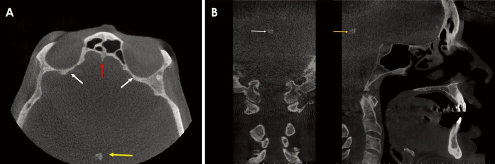
Fig. 1 A. Axial cone-beam computed tomography (CBCT) section. Arrows depict the pineal gland calcification (yellow), the crista galli (red), and the orbital cavity (white). B. Coronal (left) and sagittal (right) CBCT sections depict pineal gland calcification (white arrow in the coronal section; yellow arrow in the sagittal section).
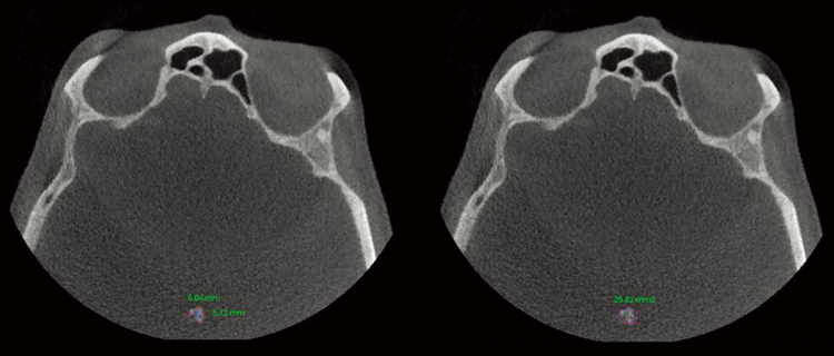
Fig. 2 Pictures depict linear (left) and areal (right) measurements of the pineal gland.
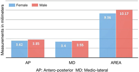
Fig. 3 Average linear and areal measurements in males and females.
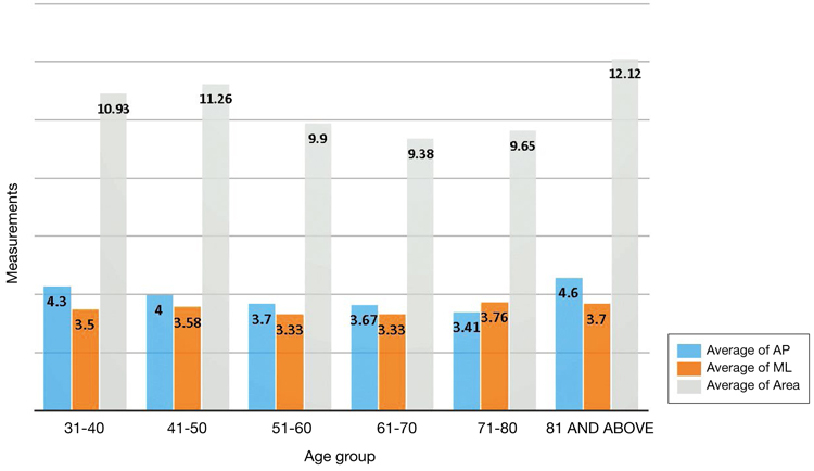
Fig. 4 Distribution of pineal gland measurements in different age groups.
Reference
-
1. Sedghizadeh PP, Nguyen M, Enciso R. Intracranial physiological calcifications evaluated with cone beam CT. Dentomaxillofac Radiol. 2012; 41:675–678.
Article2. Barghan S, Tahmasbi Arashlow M, Nair MK. Incidental findings on cone beam computed tomography studies outside of the maxillofacial skeleton. Int J Dent. 2016; 2016:9196503.
Article3. Damaskos S, Tsiklakis K, Syriopoulos K, van der Stelt P. Extra-and intra-cranial arterial calcifications in adults depicted as incidental findings on cone beam CT images. Acta Odontol Scand. 2015; 73:202–209.4. Kitkhuandee A, Sawanyawisuth K, Johns NP, Kanpittaya J, Johns J. Pineal calcification is associated with symptomatic cerebral infarction. J Stroke Cerebrovasc Dis. 2014; 23:249–253.
Article5. Mahlberg R, Walther S, Kalus P, Bohner G, Haedel S, Reischies FM, et al. Pineal calcification in Alzheimer's disease: an in vivo study using computed tomography. Neurobiol Aging. 2008; 29:203–209.
Article6. Mohammed KA, Adjei Boakye E, Ismail HA, Geneus CJ, Tobo BB, Buchanan PM, et al. Pineal gland calcification in Kurdistan: a cross-sectional study of 480 roentgenograms. PLoS One. 2016; 11:e0159239.
Article7. Bersani G, Garavini A, Taddei I, Tanfani G, Nordio M, Pancheri P. Computed tomography study of pineal calcification in schizophrenia. Eur Psychiatry. 1999; 14:163–166.
Article8. Edwards R, Altalibi M, Flores-Mir C. The frequency and nature of incidental findings in cone-beam computed tomographic scans of the head and neck region: a systematic review. J Am Dent Assoc. 2013; 144:161–170.9. Whitehead MT, Oh C, Raju A, Choudhri AF. Physiologic pineal region, choroid plexus, and dural calcifications in the first decade of life. AJNR Am J Neuroradiol. 2015; 36:575–580.
Article10. Turgut AT, Karakaş HM, Ozsunar Y, Altın L, Ceken K, Alıcıoğlu B, et al. Age-related changes in the incidence of pineal gland calcification in Turkey: a prospective multicenter CT study. Pathophysiology. 2008; 15:41–48.
Article11. Doyle AJ, Anderson GD. Physiologic calcification of the pineal gland in children on computed tomography: prevalence, observer reliability and association with choroid plexus calcification. Acad Radiol. 2006; 13:822–826.
Article12. Kunz D, Schmitz S, Mahlberg R, Mohr A, Stöter C, Wolf KJ, et al. A new concept for melatonin deficit: on pineal calcification and melatonin excretion. Neuropsychopharmacology. 1999; 21:765–772.
Article13. Ozlece HK, Akyuz O, Ilik F, Huseyinoglu N, Aydin S, Can S, et al. Is there a correlation between the pineal gland calcification and migraine? Eur Rev Med Pharmacol Sci. 2015; 19:3861–3864.14. Sandyk R. The pineal gland and the mode of onset of schizophrenia. Int J Neurosci. 1992; 67:9–17.
Article15. Wu YH, Swaab DF. The human pineal gland and melatonin in aging and Alzheimer's disease. J Pineal Res. 2005; 38:145–152.
Article16. Sigurdardottir LG, Markt SC, Sigurdsson S, Aspelund T, Fall K, Schernhammer E, et al. Pineal gland volume assessed by MRI and its correlation with 6-sulfatoxymelatonin levels among older men. J Biol Rhythms. 2016; 31:461–469.
Article17. Slominski RM, Reiter RJ, Schlabritz-Loutsevitch N, Ostrom RS, Slominski AT. Melatonin membrane receptors in peripheral tissues: distribution and functions. Mol Cell Endocrinol. 2012; 351:152–166.
Article18. Sandyk R, Anastasiadis PG, Anninos PA, Tsagas N. Is the pineal gland involved in the pathogenesis of endometrial carcinoma. Int J Neurosci. 1992; 62:89–96.
Article
- Full Text Links
-
- Actions
-
Cited
- CITED
-
- Close
- Share
-
- Similar articles
-
- Pineal calcification on computed tomography
- A Statistical Study of Pineal Body Calcification
- Pineal Gland Metastasis as the Initial Presentation of Squamous Cell Lung Cancer: A Case Report
- A case report of incidental finding of fungus ball on CBCT of maxillary sinus in treatment planning of dental implant
- Regulation of Melatonin Synthesis and Release in the pineal Gland
