Classification and management of eyelid disorders
- Affiliations
-
- 1Saevit Eye Hospital, Goyang, Korea. lkw740306@hanmail.net
- KMID: 2389792
- DOI: http://doi.org/10.5124/jkma.2017.60.9.732
Abstract
- The eyelids can be affected by various congenital, acquired, infectious, inflammatory, neoplastic, and traumatic conditions. The eyelid is not simply made up of skin, but is a complex anatomical structure. The various structures that support and protect the eyeball are gathered together in narrow spaces. Therefore, the disorders that can occur in the eyelids vary depending on the structures affected. The eyeball can be directly and indirectly influenced by each of these disorders. Thus, each eyelid disorder occurs within the frame of the corresponding anatomical structure, and must be precisely understood and treated accordingly.
Keyword
MeSH Terms
Figure
-
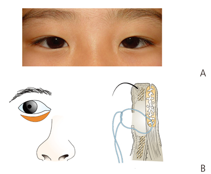
Figure 1 (A) Lower lid epiblepharon. (B) Modified Hotz's operation. Reproduced from Korean Society of Ophthalmic Plastic and Reconstructive Surgery. Ophthalmic plastic and reconstructive surgery. 3rd ed. Seoul: Naewoi Haksul; 2015, with permission from Korean Society of Ophthalmic Plastic and Reconstructive Surgery [2].
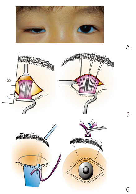
Figure 2 (A) Congenital blepharoptosis (right upper eyelid). (B) Levator resection operation. (C) Pentagonal shape frontalis sling operation. Reproduced from Korean Society of Ophthalmic Plastic and Reconstructive Surgery. Ophthalmic plastic and reconstructive surgery. 3rd ed. Seoul: Naewoi Haksul; 2015, with permission from Korean Society of Ophthalmic Plastic and Reconstructive Surgery [2].
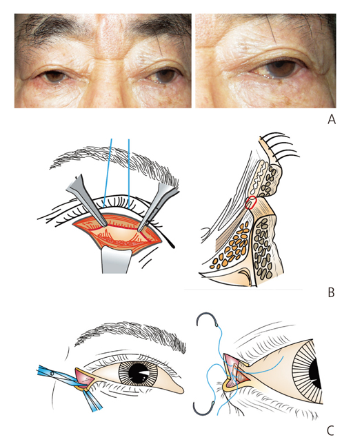
Figure 3 (A) Involutional entropion. (B) Lower lid retractor reinsertion operation. (C) Lateral tarsal strip operation. Reproduced from Korean Society of Ophthalmic Plastic and Reconstructive Surgery. Ophthalmic plastic and reconstructive surgery. 3rd ed. Seoul: Naewoi Haksul; 2015, with permission from Korean Society of Ophthalmic Plastic and Reconstructive Surgery [2].
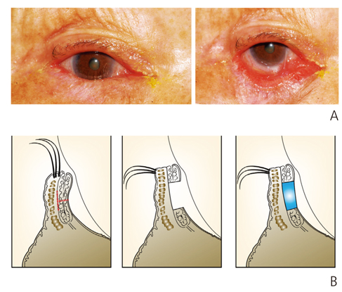
Figure 4 (A) Cicatricial entropion. (B) Posterior lamellar lengthening by tissue graft. Reproduced from Korean Society of Ophthalmic Plastic and Reconstructive Surgery. Ophthalmic plastic and reconstructive surgery. 3rd ed. Seoul: Naewoi Haksul; 2015, with permission from Korean Society of Ophthalmic Plastic and Reconstructive Surgery [2].
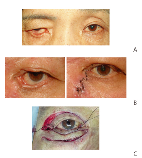
Figure 5 (A) Right lower lid ectropion. Reproduced from Korean Society of Ophthalmic Plastic and Reconstructive Surgery. Ophthalmic plastic and reconstructive surgery. 3rd ed. Seoul: Naewoi Haksul; 2015, with permission from Korean Society of Ophthalmic Plastic and Reconstructive Surgery [2]. (B) Z plasty. (C) Full thickness skin graft.

Figure 6 Levator aponeurosis repair operation. Reproduced from Korean Society of Ophthalmic Plastic and Reconstructive Surgery. Ophthalmic plastic and reconstructive surgery. 3rd ed. Seoul: Naewoi Haksul; 2015, with permission from Korean Society of Ophthalmic Plastic and Reconstructive Surgery [2].

Figure 7 Nevus. Reproduced from Korean Society of Ophthalmic Plastic and Reconstructive Surgery. Ophthalmic plastic and reconstructive surgery. 3rd ed. Seoul: Naewoi Haksul; 2015, with permission from Korean Society of Ophthalmic Plastic and Reconstructive Surgery [2].

Figure 8 Basal cell carcinoma. Reproduced from Korean Society of Ophthalmic Plastic and Reconstructive Surgery. Ophthalmic plastic and reconstructive surgery. 3rd ed. Seoul: Naewoi Haksul; 2015, with permission from Korean Society of Ophthalmic Plastic and Reconstructive Surgery [2].
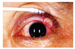
Figure 9 Squamous cell carcinoma. Reproduced from Korean Society of Ophthalmic Plastic and Reconstructive Surgery. Ophthalmic plastic and reconstructive surgery. 3rd ed. Seoul: Naewoi Haksul; 2015, with permission from Korean Society of Ophthalmic Plastic and Reconstructive Surgery [2].

Figure 10 Sebaceous cell carcinoma. Reproduced from Korean Society of Ophthalmic Plastic and Reconstructive Surgery. Ophthalmic plastic and reconstructive surgery. 3rd ed. Seoul: Naewoi Haksul; 2015, with permission from Korean Society of Ophthalmic Plastic and Reconstructive Surgery [2].
Cited by 1 articles
-
Introduction of ophthalmic plastic and reconstructive surgery
Suk-Woo Yang
J Korean Med Assoc. 2017;60(9):717-718. doi: 10.5124/jkma.2017.60.9.717.
Reference
-
1. Yi K, Ku HJ, Kim TW, Kim YD. Surgical correction for epiblepharon. J Korean Ophthalmol Soc. 1998; 39:11–16.2. Korean Society of Ophthalmic Plastic and Reconstructive Surgery. Ophthalmic plastic and reconstructive surgery. 3rd ed. Seoul: Naewoi Haksul;2015.3. Jeong SK, Park HJ, Ma YR. Surgical correction of congenital epiblepharon: lower eyelid crease reforming technique. J Korean Ophthalmol Soc. 2000; 41:8–11.4. Kim SY, Chung WS. Analysis of the causes of ptosis. J Korean Ophthalmol Soc. 1995; 36:1649–1654.5. SooHoo JR, Davies BW, Allard FD, Durairaj VD. Congenital ptosis. Surv Ophthalmol. 2014; 59:483–492.
Article6. Lee V, Konrad H, Bunce C, Nelson C, Collin JR. Aetiology and surgical treatment of childhood blepharoptosis. Br J Ophthalmo. 2002; 86:1282–1286.
Article7. Heher KL, Katowitz JA, Hollsten DA. Involutional entropion. In : Levine MR, editor. Manual of oculoplastic surgery. 2nd ed. Boston: Butterworth-Heinemann;1996. p. 119–128.8. Nerad JA. The diagnosis and treatment of entropion. In : Krachmer JH, editor. Oculoplastic surgery: the requisites in ophthalmology. 1st ed. St. Louis: Mosby;2001. p. 89–103.9. Ahn Y, Kang IS. Correction of involutional entropion by the amount of lower eyelid laxity. J Korean Ophthalmol Soc. 1999; 40:596–602.10. Anderson RL, Gordy DD. The tarsal strip procedure. Arch Ophthalmol. 1979; 97:2192–2196.
Article11. Baek SH, Kim YD. Hard palate mucosa grafts for cicatricial entropion. J Korean Ophthalmol Soc. 1996; 37:541–548.12. Nerad JA. Diagnosis and treatment of ectropion. In : Nerad JA, editor. Techniques in ophthalmic plastic surgery: a personal tutorial. Philadelphia: Saunders Elsevier;2010. p. 81–97.13. Levine MR. Involutional ectropion repair. In : Levine MR, editor. Manual of oculoplastic surgery. 3rd ed. Philadelphia: Butterworth-Heinemann;2003. p. 163–179.14. Choe KS, Kim YS, Lee TS. A clinical study of surgical results on 456 blepharoptosis. J Korean Ophthalmol Soc. 1995; 36:1093–1104.15. Allen RC, Saylor MA, Nerad JA. The current state of ptosis repair: a comparison of internal and external approaches. Curr Opin Ophthalmol. 2011; 22:394–399.16. American Academy of Ophthalmology. Orbital neoplasma. American Academy of Ophthalmology. Orbit, eyelids, and lacrimal system. San Francisco: American Academy of Ophthalmology;2008. p. 81–88.17. Bernardini FP. Management of malignant and benign eyelid lesions. Curr Opin Ophthalmol. 2006; 17:480–484.
Article18. Nemet AY, Deckel Y, Martin PA, Kourt G, Chilov M, Sharma V, Benger R. Management of periocular basal and squamous cell carcinoma: a series of 485 cases. Am J Ophthalmol. 2006; 142:293–297.
Article19. Limawararut V, Leibovitch I, Sullivan T, Selva D. Periocular squamous cell carcinoma. Clin Exp Ophthalmol. 2007; 35:174–185.
Article20. Shields JA, Demirci H, Marr BP, Eagle RC Jr, Shields CL. Sebaceous carcinoma of the eyelids: personal experience with 60 cases. Ophthalmology. 2004; 111:2151–2157.
- Full Text Links
-
- Actions
-
Cited
- CITED
-
- Close
- Share
-
- Similar articles
-
- Age-related eyelid changes
- The Psychosomatic Disorders Pertaining to Dental Practice with Revised Working Type Classification
- New Classification of Tremors: 2018 International Parkinson and Movement Disorder Society
- Epiblepharon of the lower eyelid: classification and association with astigmatism
- High-resolution Manometry: Esophageal Disorders Not Addressed by the "Chicago Classification"
