Congenital Absence of Superior Vena Cava with no Manifestation of Superior Vena Cava Syndrome
- Affiliations
-
- 1Cardiovascular Center, Seoul National University Bundang Hospital, Seongnam, Korea. jinjooparkmd@gmail.com
- 2Cardiovascular Center, Seoul National University Hospital, Seoul, Korea.
- 3Department of Radiology, Seoul National University Bundang Hostpial, Seongnam, Korea.
- KMID: 2389636
- DOI: http://doi.org/10.4070/kcj.2016.46.5.743
Abstract
- Total absence of superior vena cava (SVC) is a very rare anomaly, and the patient usually suffers from SVC syndrome or conduction disturbances. We report an asymptomatic 27 year-old male, with complete absence of SVC. Transthoracic echocardiography and computed tomography demonstrated the absence of SVC and other congenital cardiac anomalies, but the presence of prominent collateral vessels that allow a sufficient venous return.
MeSH Terms
Figure
-
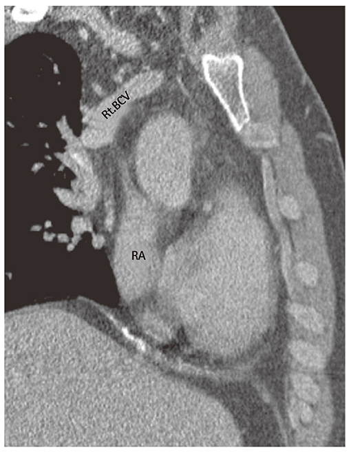
Fig. 1 Reconstructed computed tomography scan showed the absence of superior vena cava (SVC). After confluence of the brachiocephalic veins, the SVC was absent. RA: right atrium, Rt.BCV: right brachiocephalic vein.
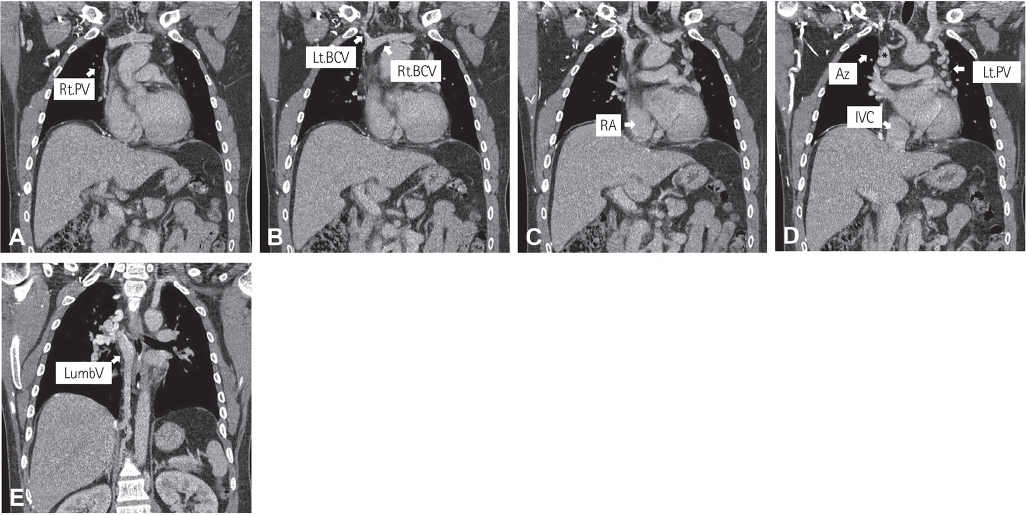
Fig. 2 (A-E) These coronal images were obtained from contrast computed tomography scan. Prominently dilated collateral vessels were observed. After confluence of left and right brachiocephalic veins, blood flow was no more visible. (C, D) Contrary to absence of SVC, blood from the IVC flows into the right atrium intact. Rt.PV: right pericardiphrenic vein, Lt.PV: left pericardiophrenic vein, RA: right atrium, Az: Azygos vein (*), IVC: inferior vena cava, LumbV: lumbar vein, SVC: superior vena cava.
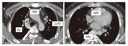
Fig. 3 (A, B) Axial images taken via contrast computed tomography. Dilatation of various collateral vessels is evident. IMV: internal mammary vein, Lt.PV: left pericardiophrenic vein, HAz: hemiazygos vein, Az: Azygos vein (*).
Reference
-
1. Wudel LJ Jr, Nesbitt JC. Superior vena cava syndrome. Curr Treat Options Oncol. 2001; 2:77–91.2. Ylänen K, Poutanen T, Savikurki-Heikkilä P, Uotila J, Korppi M, Eerola A. Bilateral absence of the superior vena cava. Case Rep Cardiol. 2012; 2012:461040.3. Wilson LD, Detterbeck FC, Yahalom J. Clinical practice. Superior vena cava syndrome with malignant causes. N Engl J Med. 2007; 356:1862–1869.4. Wan JF, Bezjak A. Superior vena cava syndrome. Hematol Oncol Clin North Am. 2010; 24:501–513.5. Bartram U, Van Praagh S, Levine JC, Hines M, Bensky AS, Van Praagh R. Absent right superior vena cava in visceroatrial situs solitus. Am J Cardiol. 1997; 80:175–183.6. Lenox CC, Zuberbuhler JR, Park SC, et al. Absent right superior vena cava with persistent left superior vena cava: implications and management. Am J Cardiol. 1980; 45:117–122.7. Romer S, Opgen-Rhein B, Chaoui R, Scheer I, Czernik C, Obladen M. Bilateral agenesis of the superior vena cava associated with congenital hydrothorax. Ultrasound Obstet Gynecol. 2006; 28:842–844.8. Lee CY, Jan SL, Wang TM, Chi CS. Congenital chylothorax associated with isolated congenital hypoplastic superior caval vein: a case report. Acta paediatr. 2005; 94:1840–1843.9. Saunders RN, Richens DR, Morris GK. Bilateral absence of the superior vena cava. Ann Thorac Surg. 2001; 71:2041–2043.
- Full Text Links
-
- Actions
-
Cited
- CITED
-
- Close
- Share
-
- Similar articles
-
- A Case of Superior Vena Cava Syndrome
- Persistent Left Superior Vena Cava with Absent Right Superior Vena Cava and Large Atrial Septal Defect in Visceroatrial Situs solitus
- Treatment of Superior Vena Cava Syndrome
- Congenital Absence of the Azygos Vein with Persistent Left Superior Vena Cava: A Case Report
- A Case of Behcet's Disease with Superior Vena Cava Syndrome
