Optimal Radial Motor Nerve Conduction Study Using Ultrasound in Healthy Adults
- Affiliations
-
- 1Department of Rehabilitation Medicine, Soonchunhyang University Cheonan Hospital, Soonchunhyang University College of Medicine, Cheonan, Korea. simon108@naver.com
- 2Department of Rehabilitation Medicine, Soonchunhyang University Bucheon Hospital, Soonchunhyang University College of Medicine, Bucheon, Korea.
- KMID: 2389488
- DOI: http://doi.org/10.5535/arm.2017.41.2.290
Abstract
OBJECTIVE
To obtain reference values, to suggest optimal recording and stimulation site for radial motor nerve conduction study (RmNCS), and to analyze the correlation among RmNCS parameters, demographics and ultrasonography (US) findings.
METHODS
A total of 55 volunteers participated in this study. We hypothesized that "˜lateral edge of spiral groove (A)' was the optimal stimulation site, and the "˜largest cross-sectional area (CSA) of extensor indicis proprius (EIP) muscle (B)' was the optimal recording site. The surface distance between "˜A' and the lateral epicondyle of the humerus divided by upper arm length, was named the spiral groove ratio. The surface distance between "˜B' and the ulnar styloid process divided by forearm length, was named the EIP ratio. Using US, we identified these sites, and further conducted RmNCS.
RESULTS
Data was collected from 100 arms of the 55 volunteers. Mean amplitude and latency were 5.7±1.1 mV and 5.7±0.5 ms, respectively, at the spiral groove, and velocity between elbow and spiral groove was 73.7±7.0 m/s. RmNCS parameters correlated significantly with height, weight, arm length, and CSA of the EIP muscle. Spiral groove ratio and EIP ratio were 0.338±0.03 and 0.201±0.03, respectively; both values were almost the same, regardless of age, sex and handedness.
CONCLUSION
We established a reference value and standardized method of RmNCS using US. Optimal RmNCS can be conducted by placing the recording electrode 20% (about one-fifth) of forearm length from the ulnar styloid process, and stimulating at 34% (about one-third) of the humeral length from the lateral epicondyle.
Keyword
MeSH Terms
Figure
-
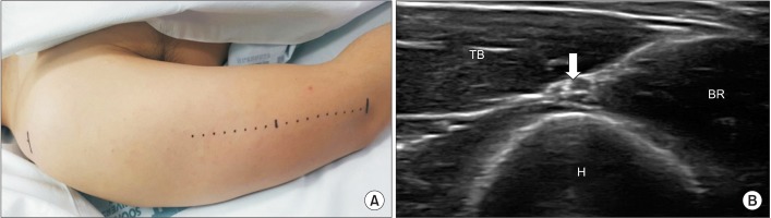
Fig. 1 Ultrasonographic evaluation of radial nerve at the spiral groove. (A) The probe was placed along the straight line between lateral epicondyle of humerus and most lateral point of acromion process of scapula. The probe was then tracked in the proximal and distal directions, until the radial nerve was located at its most superficial level above the humerus. (B) Ultrasonographic evaluation of radial nerve which placed most superficially between TB and BR at the lateral edge of spiral groove (arrow, radial nerve with brachial artery). H, humerus; TB, triceps brachii; BR, brachialis.
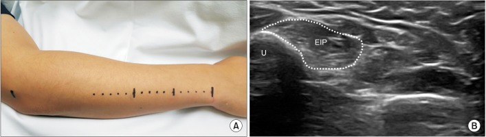
Fig. 2 Ultrasonographic evaluation of EIP muscle. (A) The probe was placed along the straight line between styloid process of ulna and lateral epicondyle of humerus. The probe was then tracked in the proximal and distal direction to find the thickest site of EIP muscle. On the basis of that site, the largest cross-sectional area (CSA) of EIP muscle was measured. To reduce error, CSA was measured twice, and the average value was calculated. (B) Ultrasonographic evaluation of EIP muscle to find largest CSA site (dotted line, CSA of EIP). EIP, extensor indicis proprius; U, ulna.
Cited by 1 articles
-
Ultrasonographic Analysis of Optimal Needle Placement for Extensor Indicis
Jin Young Kim, Hyun Seok, Sang-Hyun Kim, Yoon-Hee Choi, Jun Young Ahn, Seung Yeol Lee
Ann Rehabil Med. 2020;44(6):450-458. doi: 10.5535/arm.20035.
Reference
-
1. Dumitru D, Amato AA, Zwarts MJ. Electrodiagnostic medicine. 2nd ed. Philadelphia: Hanley & Belfus;1995. p. 810–811. p. 1087–1094.2. Kimura J. Electrodiagnosis in diseases of nerve and muscle: principles and practice. 3rd ed. New York: Oxford University Press;2001. p. 148–151. p. 717–718.3. Mondelli M, Morana P, Ballerini M, Rossi S, Giannini F. Mononeuropathies of the radial nerve: clinical and neurographic findings in 91 consecutive cases. J Electromyogr Kinesiol. 2005; 15:377–383. PMID: 15811608.
Article4. Preston DC, Shapiro BE. Electromyography and neuromuscular disorders. 3rd ed. Philadelphia: Elsevier Saunders;2013. p. 331–345.5. Lee HJ, DeLisa JA. Manual of nerve conduction study and surface anatomy for needle electromyography. Philadelphia: Lippincott Williams & Wilkins;2005. p. 55–56.6. Jebsen RH. Motor conduction velocity of distal radial nerve. Arch Phys Med Rehabil. 1966; 47:12–16. PMID: 5902993.7. Papathanasiou ES, Zamba E, Papacostas SS. Radial nerve F-waves: normative values with surface recording from the extensor indicis muscle. Clin Neurophysiol. 2001; 112:145–152. PMID: 11137672.
Article8. Kang SY, Ko YJ, Choi ES, Oh CS. Radial motor nerve conduction study using surface electrode in normal adults. J Korean Acad Rehabil Med. 1992; 16:385–389.9. Trojaborg W, Sindrup EH. Motor and sensory conduction in different segments of the radial nerve in normal subjects. J Neurol Neurosurg Psychiatry. 1969; 32:354–359. PMID: 5807873.
Article10. Boon AJ, Bailey PW, Smith J, Sorenson EJ, Harper CM, Hurdle MF. Utility of ultrasound-guided surface electrode placement in lateral femoral cutaneous nerve conduction studies. Muscle Nerve. 2011; 44:525–530. PMID: 21826680.
Article11. Kamm CP, Scheidegger O, Rosler KM. Ultrasound-guided needle positioning in sensory nerve conduction study of the sural nerve. Clin Neurophysiol. 2009; 120:1342–1345. PMID: 19464944.
Article12. Oh CH, Park NS, Kim JM, Kim MW. Determination of an ideal stimulation site of the medial antebrachial cutaneous nerve using ultrasound and investigation of the efficiency. Ann Rehabil Med. 2014; 38:836–842. PMID: 25566484.
Article13. Park BJ, Joeng ES, Choi JK, Kang S, Yoon JS, Yang SN. Ultrasound-guided lateral femoral cutaneous nerve conduction study. Ann Rehabil Med. 2015; 39:47–51. PMID: 25750871.
Article14. Boon AJ, Alsharif KI, Harper CM, Smith J. Ultrasound-guided needle EMG of the diaphragm: technique description and case report. Muscle Nerve. 2008; 38:1623–1626. PMID: 19016552.
Article15. Lee SM, Kim K, Lee SM, Lee HS. Ultrasonographic evaluation of needle insertion site for the flexor pollicis longus. Ann Rehabil Med. 2013; 37:215–220. PMID: 23705116.
Article16. Yang SN, Lee SH, Kwon HK. Needle electrode insertion into the tibialis posterior: a comparison of the anterior and posterior approaches. Arch Phys Med Rehabil. 2008; 89:1816–1818. PMID: 18760169.
Article17. Robinson LR, Rubner DE, Wahl PW, Fujimoto WY, Stolov WC. Influences of height and gender on normal nerve conduction studies. Arch Phys Med Rehabil. 1993; 74:1134–1138. PMID: 8239949.18. Stetson DS, Albers JW, Silverstein BA, Wolfe RA. Effects of age, sex, and anthropometric factors on nerve conduction measures. Muscle Nerve. 1992; 15:1095–1104. PMID: 1406766.
Article19. Won SJ, Kim BJ, Park KS, Yoon JS, Choi H. Reference values for nerve ultrasonography in the upper extremity. Muscle Nerve. 2013; 47:864–871. PMID: 23625758.
Article20. Brown JM, Yablon CM, Morag Y, Brandon CJ, Jacobson JA. US of the peripheral nerves of the upper extremity: a landmark approach. Radiographics. 2016; 36:452–463. PMID: 26963456.
Article21. Vergara-Amador E, Ramirez A. Anatomic study of the extensor carpi radialis brevis in its relation with the motor branch of the radial nerve. Orthop Traumatol Surg Res. 2015; 101:909–912. PMID: 26547256.
Article22. Hackl M, Damerow D, Leschinger T, Scaal M, Muller LP, Wegmann K. Radial nerve location at the posterior aspect of the humerus: an anatomic study of 100 specimens. Arch Orthop Trauma Surg. 2015; 135:1527–1532. PMID: 26254580.
Article23. Fleming P, Lenehan B, Sankar R, Folan-Curran J, Curtin W. One-third, two-thirds: relationship of the radial nerve to the lateral intermuscular septum in the arm. Clin Anat. 2004; 17:26–29. PMID: 14695584.
Article24. Cauldwell EW, Anson BJ, Wright RR. The extensor indicis proprius muscle: a study of 263 consecutive specimans. Q Bull Northwest Univ Med Sch. 1943; 17:267–279.
- Full Text Links
-
- Actions
-
Cited
- CITED
-
- Close
- Share
-
- Similar articles
-
- Pitfalls in Superficial Radial Sensory Nerve Conduction Study
- A Study of Nerve Conduction Velocity of Normal Adults
- A Study of Motor Conduction Velocity of Radial Nerve: Comparision of Proximal and Distal Segments
- Nerve length measurement method in a radial motor nerve conduction study
- Radial motor nerve conduction study using surface electrode in normal adults
