Ann Rehabil Med.
2017 Jun;41(3):516-517. 10.5535/arm.2017.41.3.516.
Recovery of an Injured Corticoreticulospinal Tract in a Patient With Cerebral Infarct
- Affiliations
-
- 1Department of Physical Medicine and Rehabilitation, Yeungnam University College of Medicine, Daegu, Korea. soyoung.kwak@daum.net
- KMID: 2389471
- DOI: http://doi.org/10.5535/arm.2017.41.3.516
Abstract
- No abstract available.
MeSH Terms
Figure
-
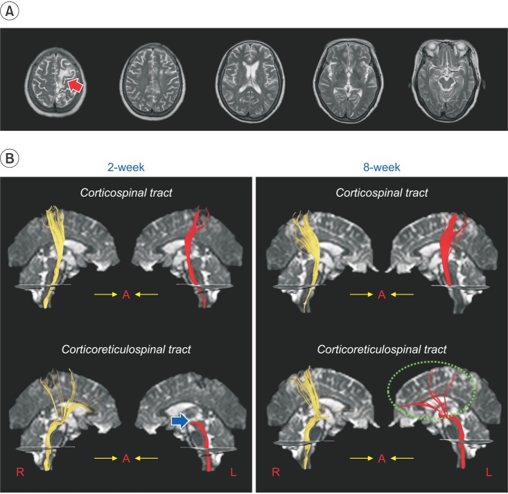
Fig. 1 (A) Brain MR images at 2 weeks after onset shows leukomalactic lesion (red arrow) in the left supplementary motor area and premotor cortex. (B) Results of diffusion tensor tractography (DTT) for the corticospinal tract (CST) and the corticoreticulospinal tract (CRT) of the patient. The 2-week DTT shows the integrity of both the CSTs and the right CRT of the patient are preserved. However, the left CRT shows discontinuation (blue arrow) at the brainstem level. By contrast, the 8-week DTT reveals the discontinued left CRT is connected to the left cerebral cortex (dotted circle).
Reference
-
1. Yeo SS, Jang SH. Recovery of an injured corticospinal tract and an injured corticoreticular pathway in a patient with intracerebral hemorrhage. NeuroRehabilitation. 2013; 32:305–309. PMID: 23535792.
Article2. Jang SH, Yeo SS. Recovery of an injured corticoreticular pathway via transcallosal fibers in a patient with intracerebral hemorrhage. BMC Neurol. 2014; 14:108. PMID: 24886278.
Article
- Full Text Links
-
- Actions
-
Cited
- CITED
-
- Close
- Share
-
- Similar articles
-
- Which Neural Tract Plays a Major Role in Memory Impairment After Multiple Cerebral Infarcts? A Case Report
- Correlation between Body Temperature and Infarct Size and Recovery in the Stroke
- A Case of Middle Cerebral Artery Infarct Developed Immediately After Head Injury
- The Comparison of Videofluoroscopic Findings between the Patients with Lateral Medullary Infarct and Middle Cerebral Artery Territorial Infarct
- Visual recovery demonstrated by functional MRI and diffusion tensor tractography in bilateral occipital lobe infarction
