Ann Dermatol.
2017 Oct;29(5):621-625. 10.5021/ad.2017.29.5.621.
Primary Cutaneous Endometriosis of Umbilicus
- Affiliations
-
- 1Department of Dermatology, Kyung Hee University College of Medicine, Seoul, Korea. bellotte@hanmail.net
- KMID: 2388916
- DOI: http://doi.org/10.5021/ad.2017.29.5.621
Abstract
- Cutaneous endometriosis is defined by the presence of endometrial glands and/or stroma in skin and represents less than 1% of all ectopic endometrium. Cutaneous endometriosis is classified as primary and secondary. Primary cutaneous endometriosis appears without a prior surgical history and secondary cutaneous endometriosis mostly occurs at surgical scar tissue after abdominal operations. The most widely accepted pathogenesis of secondary endometriosis is the iatrogenic implantation of endometrial cells after surgery, such as laparoscopic procedures. However, the pathogenesis of primary endometriosis is still unknown. Umbilical endometriosis is composed only 0.4% to 4.0% of all endometriosis, however, umbilicus is the most common site of primary cutaneous endometriosis. A 38-year-old women presented with solitary 2.5×2.0-cm-sized purple to brown colored painful nodule on the umbilicus since 2 years ago. The patient had no history of surgical procedures. The skin lesion became swollen with spontaneous bleeding during menstruation. The skin lesion was diagnosed as a keloid at private hospital and has been treated with lesional injection of steroid for several times but there was no improvement. Imaging studies showed an enhancing umbilical mass without connection to internal organs. Biopsy specimen showed the several dilated glandular structures in dermis. They were surrounded by endometrial-type stroma and perivascular infiltration of lymphocytes. The patient was diagnosed as primary cutaneous endometriosis and skin lesion was removed by complete wide excision without recurrence. We report an interesting and rare case of primary umbilical endometriosis mistaken for a keloid and review the literatures.
Keyword
MeSH Terms
Figure
-
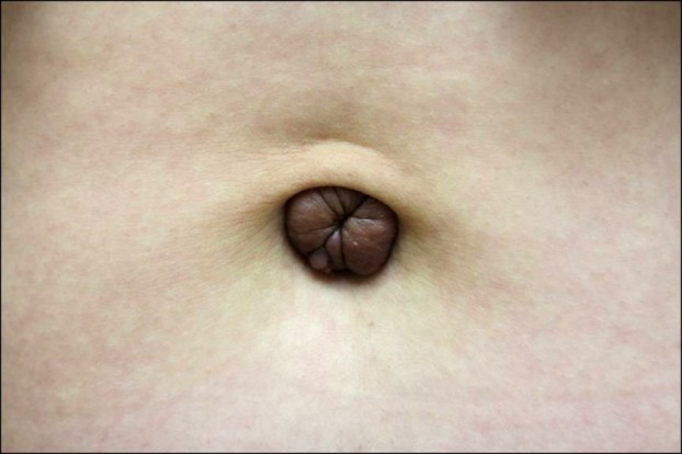
Fig. 1 About 2.5×2.0-cm-sized brownish to purple colored nodule on the umbilicus.
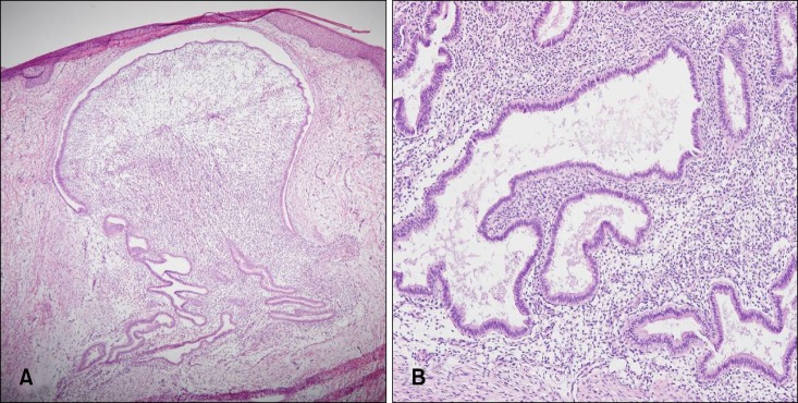
Fig. 2 (A, B) Specimen showed lesions in the superficial dermis and deep dermis comprising dilated glandular structures, surrounded by cellular endometrial-type stroma (H&E; A: ×40, B: ×100, respectively).
Reference
-
1. Minaidou E, Polymeris A, Vassiliou J, Kondi-Paphiti A, Karoutsou E, Katafygiotis P, et al. Primary umbilical endometriosis: case report and literature review. Clin Exp Obstet Gynecol. 2012; 39:562–564. PMID: 23444772.2. Yuen JS, Chow PK, Koong HN, Ho JM, Girija R. Unusual sites (thorax and umbilical hernial sac) of endometriosis. J R Coll Surg Edinb. 2001; 46:313–315. PMID: 11697703.3. Jaime TJ, Jaime TJ, Ormiga P, Leal F, Nogueira OM, Rodrigues N. Umbilical endometriosis: report of a case and its dermoscopic features. An Bras Dermatol. 2013; 88:121–124. PMID: 23539017.
Article4. Fernández-Aceñero MJ, Córdova S. Cutaneous endometriosis: review of 15 cases diagnosed at a single institution. Arch Gynecol Obstet. 2011; 283:1041–1044. PMID: 20422419.
Article5. Weng CS, Yang YC. Images in clinical medicine. Villar's nodule--umbilical endometriosis. N Engl J Med. 2011; 364:e45. PMID: 21612465.6. Victory R, Diamond MP, Johns DA. Villar's nodule: a case report and systematic literature review of endometriosis externa of the umbilicus. J Minim Invasive Gynecol. 2007; 14:23–32. PMID: 17218225.
Article7. Groothuis PG, Koks CA, de Goeij AF, Dunselman GA, Arends JW, Evers JL. Adhesion of human endometrium to the epithelial lining and extracellular matrix of amnion in vitro: an electron microscopic study. Hum Reprod. 1998; 13:2275–2281. PMID: 9756310.
Article8. Dadhwal V, Gupta B, Dasgupta C, Shende U, Deka D. Primary umbilical endometriosis: a rare entity. Arch Gynecol Obstet. 2011; 283(Suppl 1):119–120. PMID: 21170542.
Article9. Theunissen CI, IJpma FF. Primary umbilical endometriosis: a cause of a painful umbilical nodule. J Surg Case Rep. 2015; 2015:rjv025. PMID: 25786440.
Article10. Calagna G, Perino A, Chianetta D, Vinti D, Triolo MM, Rimi C, et al. Primary umbilical endometrioma: analyzing the pathogenesis of endometriosis from an unusual localization. Taiwan J Obstet Gynecol. 2015; 54:306–312. PMID: 26166347.
Article11. Chikazawa K, Mitsushita J, Netsu S, Konno R. Surgical excision of umbilical endometriotic lesions with laparoscopic pelvic observation is the way to treat umbilical endometriosis. Asian J Endosc Surg. 2014; 7:320–322. PMID: 25354378.
Article12. Pariza G, Mavrodin CI. Primary umbilical endometriosis (Villar's nodule)-case study, literature revision. Chirurgia (Bucur). 2014; 109:546–549. PMID: 25149622.13. Paramythiotis D, Stavrou G, Panidis S, Panagiotou D, Chatzopoulos K, Papadopoulos VN, et al. Concurrent appendiceal and umbilical endometriosis: a case report and review of the literature. J Med Case Rep. 2014; 8:258. PMID: 25052818.
Article14. Ghosh A, Das S. Primary umbilical endometriosis: a case report and review of literature. Arch Gynecol Obstet. 2014; 290:807–809. PMID: 24930115.
Article15. Gin TJ, Gin AD, Gin D, Pham A, Cahill J. Spontaneous cutaneous endometriosis of the umbilicus. Case Rep Dermatol. 2013; 5:368–372. PMID: 24516408.
Article16. Kahlenberg LK, Laskey S. Primary umbilical endometriosis presenting as umbilical drainage in a nulliparous and surgically naive young woman. Am J Emerg Med. 2014; 32:692.e1–692.e2.
Article17. Fancellu A, Pinna A, Manca A, Capobianco G, Porcu A. Primary umbilical endometriosis. Case report and discussion on management options. Int J Surg Case Rep. 2013; 4:1145–1148. PMID: 24291679.
Article18. Efremidou EI, Kouklakis G, Mitrakas A, Liratzopoulos N, Polychronidis ACh. Primary umbilical endometrioma: a rare case of spontaneous abdominal wall endometriosis. Int J Gen Med. 2012; 5:999–1002. PMID: 23271917.
Article19. Kesici U, Yenisolak A, Kesici S, Siviloglu C. Primary cutaneous umbilical endometriosis. Med Arch. 2012; 66:353–354. PMID: 23097979.
Article20. Bagade PV, Guirguis MM. Menstruating from the umbilicus as a rare case of primary umbilical endometriosis: a case report. J Med Case Rep. 2009; 3:9326. PMID: 20062755.
Article21. Boesgaard-Kjer D, Boesgaard-Kjer D, Kjer JJ. Primary umbilical endometriosis (PUE). Eur J Obstet Gynecol Reprod Biol. 2017; 209:44–45. PMID: 27374811.
Article22. Wiegratz I, Kissler S, Engels K, Strey C, Kaufmann M. Umbilical endometriosis in pregnancy without previous surgery. Fertil Steril. 2008; 90:199.e17–199.e20.
Article23. Taniguchi F, Hirakawa E, Azuma Y, Uejima C, Ashida K, Harada T. Primary umbilical endometriosis: unusual and rare clinical presentation. Case Rep Obstet Gynecol. 2016; 2016:9302376. PMID: 27242939.
Article24. Chew KT, Norsaadah S, Suraya A, Hing EY, Ani Amelia Z, Nor Azlin MI, et al. Primary umbilical endometriosis successfully treated with dienogest. Horm Mol Biol Clin Investig. 2017; 29:67–69.25. Claas-Quax MJ, Ooft ML, Hoogwater FJ, Veersema S. Primary umbilical endometriosis. Eur J Obstet Gynecol Reprod Biol. 2015; 194:260–261. PMID: 26344784.
Article26. Sidani MS, Khalil AM, Tawil AN, El-Hajj MI, Seoud MA. Primary umbilical endometriosis. Clin Exp Obstet Gynecol. 2002; 29:40–41. PMID: 12013091.27. Sengupta M, Naskar A, Gon S, Majumdar B. Villar's nodule. Online J Health Allied Sci. 2011; 10:19.28. Kim SH, Park SJ, Lee DY, Lee ES. A case of cutaneous endometriosis. Korean J Dermatol. 2002; 40:100–102.29. Song WK, Park HJ, Kim YC, Cinn YW. A case of cutaneous endometriosis. Korean J Dermatol. 2000; 38:999–1001.30. Kyamidis K, Lora V, Kanitakis J. Spontaneous cutaneous umbilical endometriosis: report of a new case with immunohistochemical study and literature review. Dermatol Online J. 2011; 17:5.
Article31. Purvis RS, Tyring SK. Cutaneous and subcutaneous endometriosis. Surgical and hormonal therapy. J Dermatol Surg Oncol. 1994; 20:693–695. PMID: 7930017.
