Accessory Devices Frequently Used for Endoscopic Submucosal Dissection
- Affiliations
-
- 1Division of Gastroenterology and Hepatology, Department of Internal Medicine, Institute of Gastrointestinal Medical Instrument Research, Korea University College of Medicine, Korea University Anam Hospital, Seoul, Korea. drchunhj@chol.com
- KMID: 2388818
- DOI: http://doi.org/10.5946/ce.2017.070
Abstract
- Endoscopic submucosal dissection (ESD) is increasingly being considered an essential component of treatment for early gastrointestinal cancers and subepithelial tumors. The ESD technique owes its popularity to the development of sophisticated instruments used for ESD. With an increase in the number of ESD procedures performed, there is rapid development in the number and types of endoscopic accessory devices used for such procedures. Despite the large numbers of new devices developed and marketed, the use of ESD instruments and accessory devices is largely determined by individual preferences and experiences. Accessory devices frequently used during ESD are important tools for ESD techniques. Each instrument possesses characteristic advantages and disadvantages associated with its use, and no one instrument is superior in all respects to others. In this article, we review the characteristics of endoscopic electrical knives, cap and hood, and hemostatic devices commonly used in ESD.
Figure
-
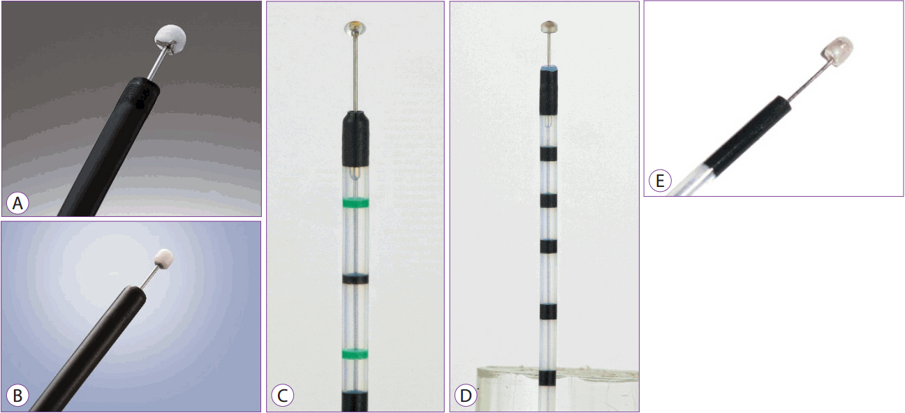
Fig. 1. (A) Insulation-tipped (IT) knife 2 (Olympus, Tokyo, Japan). (B) IT knife nano (Olympus, Tokyo, Japan). (C) Ball endoscopic submucosal dissection (ESD) knife (MTW, Wesel, Germany). (D) Hemispherical ESD knife (MTW, Wesel, Germany). (E) Helmet snare ESD knife (Kachu Technology, Seoul, Korea).
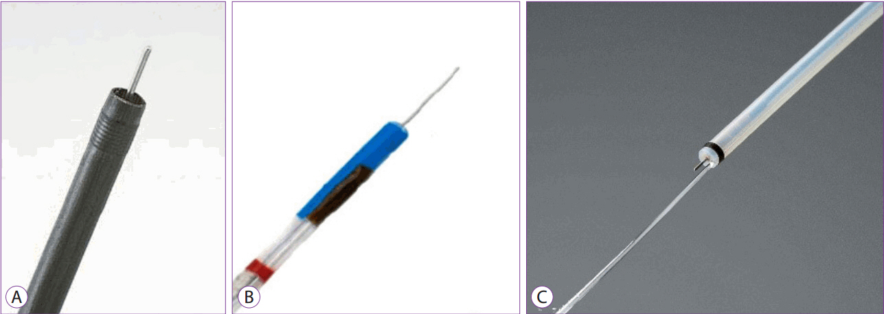
Fig. 2. (A) Needle knife (Olympus, Tokyo, Japan). (B) Needle controllable knife (MTW, Wesel, Germany). (C) Splash knife (Pentax, Tokyo, Japan).

Fig. 3. Triangle-tipped (TT) knife (Olympus, Tokyo, Japan).
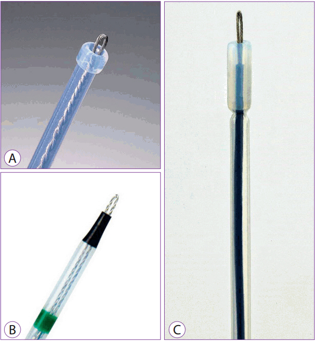
Fig. 4. (A) Flex knife (Olympus, Tokyo, Japan). (B) Fixed flexible snare knife (Kachu Technology, Seoul, Korea). (C) Universal knife (MTW, Wesel, Germany).
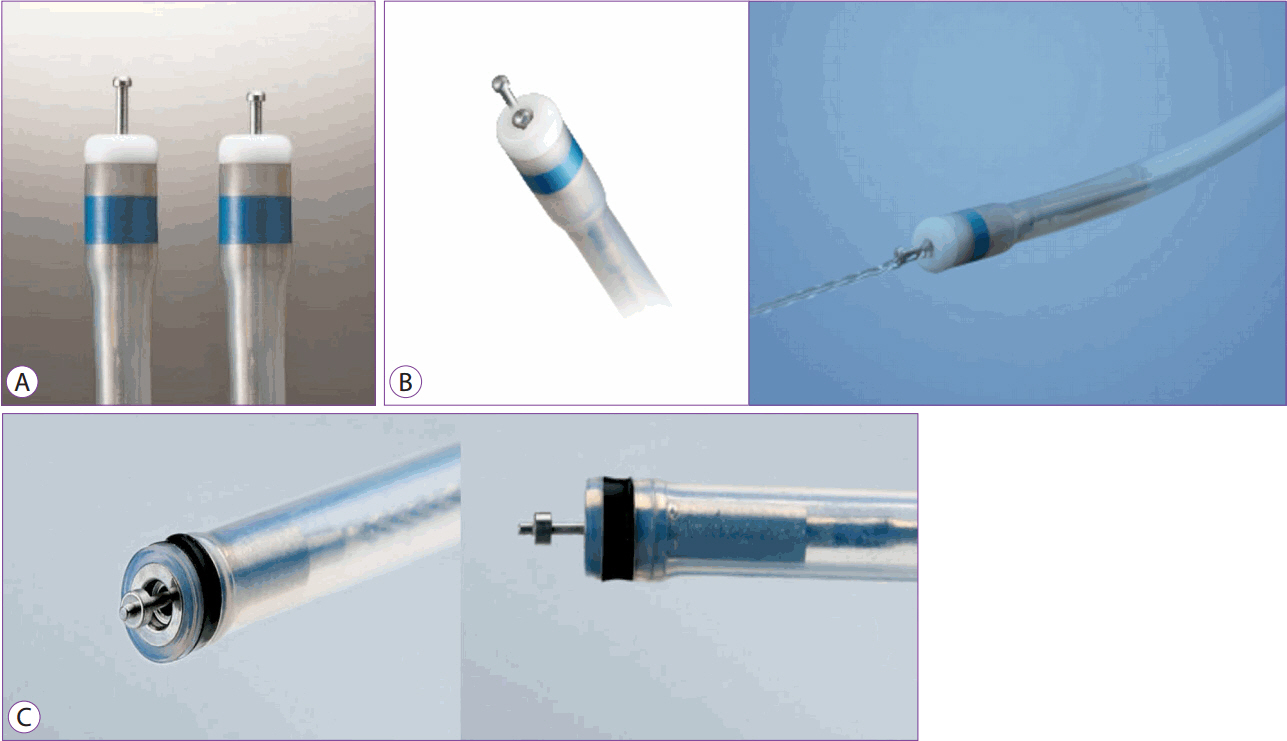
Fig. 5. (A) Dual knife (Olympus, Tokyo, Japan). (B) Dual knife J (water jet, Olympus, Tokyo, Japan). (C) Splash M knife (Pentax, Tokyo, Japan).

Fig. 6. (A) Hook knife (rotatable, Olympus, Tokyo, Japan). (B) Hook knife (MTW, Wesel, Germany).
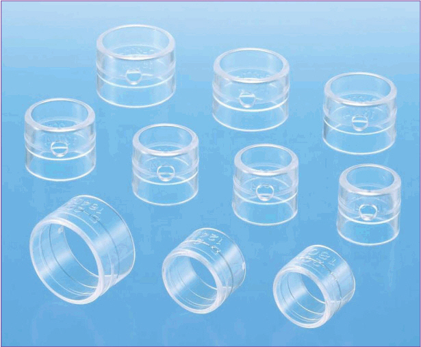
Fig. 7. Distal attachment (Olympus, Tokyo, Japan).
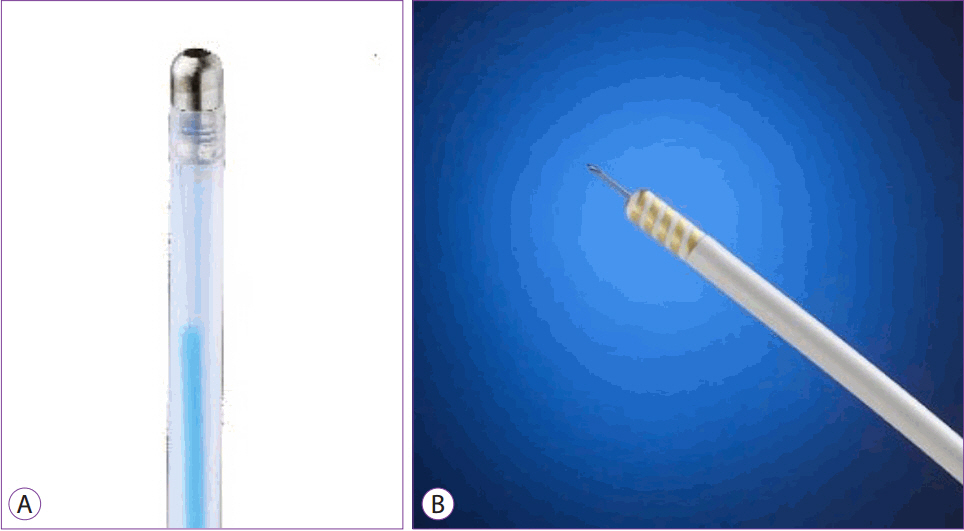
Fig. 8. (A) Coagulation probe (MTW, Wesel, Germany). (B) Injection Gold Probe TM (Boston Scientific, Marlborough, MA, USA).

Fig. 9. (A) Coagrasper (Olympus, Tokyo, Japan). (B) Hot biopsy forceps (Radial JawTM 4; Boston Scientific, Marlborough, MA, USA). (C) Hemostat-Y (Pentax, Tokyo, Japan).
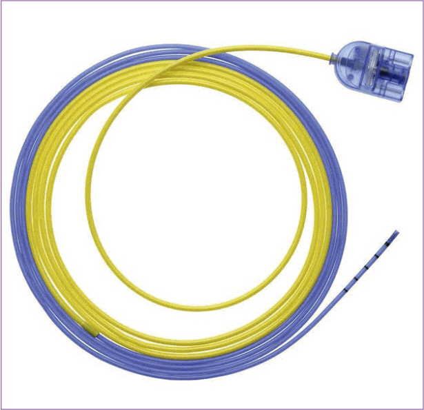
Fig. 10. Argon plasma coagulation probe (ERBE, Tübingen, Germany).
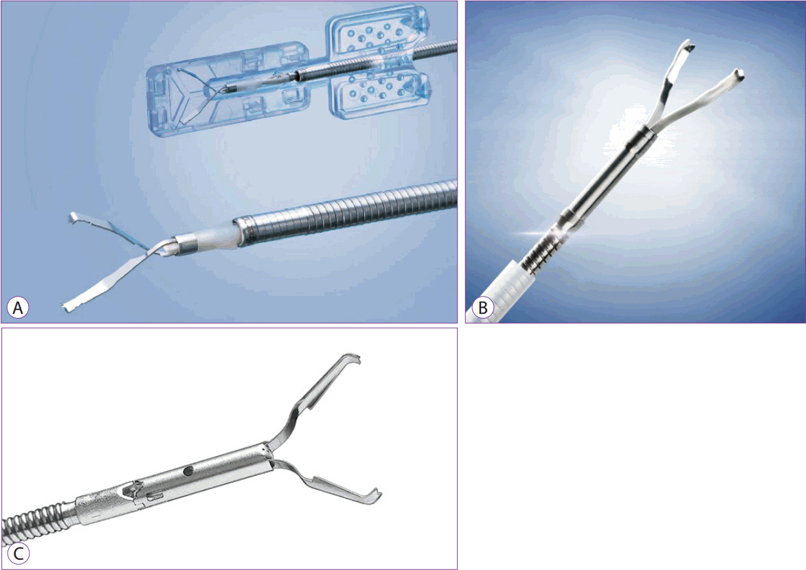
Fig. 11. (A) EZ Clip (Olympus, Tokyo, Japan). (B) QuickPro (Olympus, Tokyo, Japan). (C) Resolution clip (Boston Scientific, Marlborough, MA, USA).
Cited by 2 articles
-
Successful Endoscopic Resection of Residual Colonic Mucosa-Associated Lymphoid Tissue Lymphoma after Polypectomy
Jeongmin Choi
Clin Endosc. 2021;54(5):759-762. doi: 10.5946/ce.2020.233.Comparison between a novel core knife and the conventional IT knife 2 for endoscopic submucosal dissection of gastric mucosal lesions
Myeongsoon Park, Jin Wook Lee, Dong Woo Shin, Jungseok Kim, Yoo Jin Lee, Ju Yup Lee, Kwang Bum Cho
Clin Endosc. 2022;55(6):767-774. doi: 10.5946/ce.2022.002.
Reference
-
1. Choi HS, Kim KO, Chun HJ, et al. The efficacy of transdermal fentanyl for pain relief after endoscopic submucosal dissection: a prospective, randomised controlled trial. Dig Liver Dis. 2012; 44:925–929.
Article2. Choi HS, Chun HJ, Kim KO, et al. Endoscopic en bloc resection of an exophytic gastrointestinal stromal tumor with suction excavation technique. World J Gastroenterol. 2016; 22:5454–5458.3. Korean EUS Study Group of Korean Society of Gastrointestinal Endoscopy. Practice of endoscopic treatment for digestive tract tumor. Seoul: Medbook;2009. p. 619.4. Mori G, Nonaka S, Oda I, et al. Novel strategy of endoscopic submucosal dissection using an insulation-tipped knife for early gastric cancer: near-side approach method. Endosc Int Open. 2015; 3:E425–E431.
Article5. Suzuki T, Hara T, Kitagawa Y, Yamaguchi T. Usefulness of IT knife nano for endoscopic submucosal dissection of large colo-rectal lesions. Acta Gastroenterol Belg. 2016; 79:186–190.6. Choi HS, Chun HJ, Kim I. An unusual submucosal tumor of the stomach. Gastroenterology. 2012; 142:e11–e12.
Article7. Minami H, Isomoto H, Yamaguchi N, et al. Peroral endoscopic myotomy for esophageal achalasia: clinical impact of 28 cases. Dig Endosc. 2014; 26:43–51.8. Choi HS, Chun HJ, Seo MH, et al. Endoscopic submucosal tunnel dissection salvage technique for ulcerative early gastric cancer. World J Gastroenterol. 2014; 20:9210–9214.9. Choi HS, Keum B, Jeen YT, Chun HJ. Complete en bloc resection of an adenocarcinoma involving Brunner’s gland by using endoscopic submucosal dissection. Dig Liver Dis. 2013; 45:e7.
Article10. Soehendra N, Sriram PV, Ponchon T, Chung SC. Hemostatic clip in gastrointestinal bleeding. Endoscopy. 2001; 33:172–180.
Article11. Romagnuolo J. Endoscopic clips: past, present and future. Can J Gastroenterol. 2009; 23:158–160.
Article
- Full Text Links
-
- Actions
-
Cited
- CITED
-
- Close
- Share
-
- Similar articles
-
- History and Development of Accessories for Endoscopic Submucosal Dissection
- Debates on Colorectal Endoscopic Submucosal Dissection - Traction for Effective Dissection: Gravity Is Enough
- Future Development of Endoscopic Accessories for Endoscopic Submucosal Dissection
- A Case of Pneumorrhachis and Pneumoscrotum Following Colon Endoscopic Submucosal Dissection
- Recent Development of Techniques and Devices in Colorectal Endoscopic Submucosal Dissection
