J Korean Ophthalmol Soc.
2017 Aug;58(8):968-973. 10.3341/jkos.2017.58.8.968.
The Correlation between Cognitive Function and Glaucoma
- Affiliations
-
- 1Department of Ophthalmology, Guri Hospital, Hanyang University College of Medicine, Guri, Korea. goddns76@hanmail.net
- KMID: 2388186
- DOI: http://doi.org/10.3341/jkos.2017.58.8.968
Abstract
- PURPOSE
To compare mini-mental state examination (MMSE) score between glaucoma group and normal control group and to evaluate the correlation between MMSE score and spectral domain-optical coherence tomography (SD-OCT) values in both groups.
METHODS
This prospective study includes thirty glaucoma patients (eleven primary open angle glaucoma and nineteen normal tension glaucoma) and thirty normal controls. Retinal nerve fiber layer (RNFL) and Ganglion cell-inner plexiform layer (GC-IPL) thickness were measured with SD-OCT, and the average values of both eyes were used. The cognitive function was evaluated with MMSE by a single examiner.
RESULTS
The mean MMSE scores of glaucoma group and normal group were 26.07 ± 2.95, and 27.00 ± 1.68 respectively (p = 0.137). MMSE score of less than 24 only showed in glaucoma group. MMSE score and RNFL thickness showed statistically no signifance in correlation (R² = 0.236; p = 0.070), however, MMSE score and GC-IPL showed statistically significant correlation (R² = 0.256; p = 0.048).
CONCLUSIONS
Glaucoma patients tend to show low cognitive function even though the correlation between glaucoma patient and low cognitive function was not statistically significant. Therefore, the aspect of cognitive depression should be concerned, when facing glaucoma patients.
MeSH Terms
Figure
-
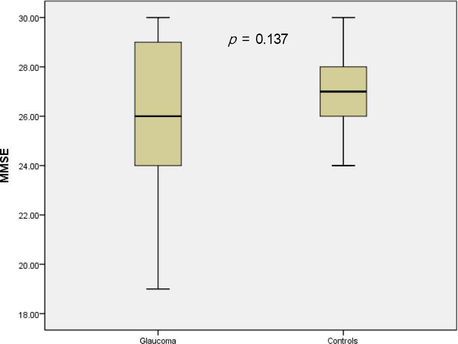
Figure 1. Box plot graph of mini-mental state examination (MMSE) scores in glaucoma and control groups. The mean score of MMSE was 26.07 ± 2.95 in glaucoma group, and 27.00 ± 1.68 in normal control group, respectively. Glaucoma group showed low MMSE score than normal con-trol group, but it was not statistically significant.
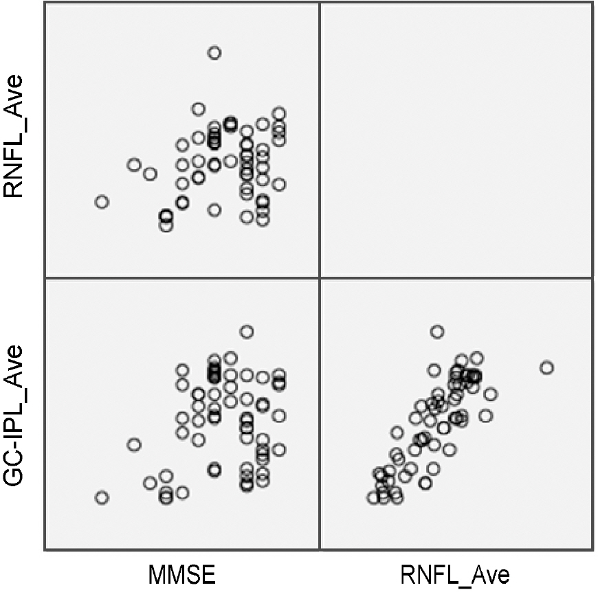
Figure 2. Scatter plot of mini-mental state examination (MMSE) score and spectral domain– optical coherence tomography (SD-OCT) parameters. Scatter plot shows weak correlation be-tween MMSE score and SD-OCT parameters. RNFL = retinal nerve fiber layer; GC-IPL = ganglion cell-inner plexiform layer.
Reference
-
References
1. Resnikoff S, Pascolini D, Etya'ale D. . Global data on visual impairment in the year 2002. Bull World Health Organ. 2004; 82:844–51.2. Kim CS, Seong GJ, Lee NH. . Prevalence of primary open-angle glaucoma in central South Korea the Namil study. Ophthalmology. 2011; 118:1024–30.3. Ferri CP, Prince M, Brayne C. . Global prevalence of dementia: a Delphi consensus study. Lancet. 2005; 366:2112–7.
Article4. Mandas A, Mereu RM, Catte O. . Cognitive impairment and age-related vision disorders: their possible relationship and the evaluation of the use of aspirin and statins in a 65 years-and-over Sardinian population. Front Aging Neurosci. 2014; 6:309.
Article5. Lin IC, Wang YH, Wang TJ. . Glaucoma, Alzheimer’s disease, and Parkinson’s disease: an 8-year population-based follow-up study. PLoS One. 2014; 9:e108938.
Article6. Shiose Y, Kitazawa Y, Tsukahara S. . Epidemiology of glauco-ma in Japan–a nationwide glaucoma survey. Jpn J Ophthalmol. 1991; 35:133–55.7. Henderson VW. The epidemiology of estrogen replacement ther-apy and Alzheimer’s disease. Neurology. 1997; 48((5 Suppl 7)):S27–35.
Article8. Sadun AA, Bassi CJ. Optic nerve damage in Alzheimer’s disease. Ophthalmology. 1990; 97:9–17.
Article9. McKinnon SJ, Lehman DM, Kerrigan-Baumrind LA. . Caspase activation and amyloid precursor protein cleavage in rat ocular hypertension. Invest Ophthalmol Vis Sci. 2002; 43:1077–87.10. Bayer AU, Ferrari F, Erb C. High occurrence rate of glaucoma among patients with Alzheimer’s disease. Eur Neurol. 2002; 47:165–8.
Article11. Yochim BP, Mueller AE, Kane KD, Kahook MY. Prevalence of cognitive impairment, depression, and anxiety symptoms among older adults with glaucoma. J Glaucoma. 2012; 21:250–4.
Article12. Kessing LV, Lopez AG, andersen PK, Kessing SV. No increased risk of developing Alzheimer disease in patients with glaucoma. J Glaucoma. 2007; 16:47–51.
Article13. Tamura H, Kawakami H, Kanamoto T. . High frequency of open-angle glaucoma in Japanese patients with Alzheimer’s disease. J Neurol Sci. 2006; 246:79–83.
Article14. Jefferis JM, Taylor JP, Collerton J. . The association between diagnosed glaucoma and cataract and cognitive performance in very old people: cross-sectional findings from the newcastle 85+ study. Ophthalmic Epidemiol. 2013; 20:82–8.
Article15. Cumurcu T, Cumurcu BE, Celikel FC, Etikan I. Depression and anxiety in patients with pseudoexfoliative glaucoma. Gen Hosp Psychiatry. 2006; 28:509–15.
Article16. Hagerman KE, Taussig MJ, Coalter JD, Jay WM. Low-vision re-habilitation in patients with visual and cognitive impairment. Vis Impair Res. 2007; 9:19–22.
Article17. Shen Y, Shi Z, Jia R. . The attenuation of retinal nerve fiber layer thickness and cognitive deterioration. Front Cell Neurosci. 2013; 7:142.
Article18. Oktem EO, Derle E, Kibaroglu S. . The relationship between the degree of cognitive impairment and retinal nerve fiber layer thickness. Neurol Sci. 2015; 36:1141–6.
Article19. Bayhan HA, Aslan Bayhan S, Celikbilek A. . Evaluation of the chorioretinal thickness changes in Alzheimer’s disease using spectraldomain optical coherence tomography. Clin Experiment Ophthalmol. 2015; 43:145–51.
Article20. Gharbiya M, Trebbastoni A, Parisi F. . Choroidal thinning as a new finding in Alzheimer’s disease: evidence from enhanced depth imaging spectral domain optical coherence tomography. J Alzheimers Dis. 2014; 40:907–17.
Article
- Full Text Links
-
- Actions
-
Cited
- CITED
-
- Close
- Share
-
- Similar articles
-
- Effects of Depression and Anxiety Symptoms on Specific Cognitive Function by Evaluating Healthy Subjects
- Correlation between Cognitive Functions and Psychotic Symptoms in Schizophrenic Patients
- Relations between Somatic Symptoms, Depression, Anxiety, and Cognitive Function in Patients with Mild Traumatic Brain Injury
- Relationships between Psychotic Symptoms and Cognitive Functions in Schizophrenic Patients
- Characteristics of Cognitive Impairment in Patients With Post-stroke Aphasia
