Fixed prosthetic treatment for the patient with delayed eruption disorder
- Affiliations
-
- 1Department of Prothodontics, School of Dentistry, Chosun University, Gwangju, Republic of Korea. khjdds@chosun.ac.kr
- KMID: 2388019
- DOI: http://doi.org/10.14368/jdras.2017.33.2.127
Abstract
- Delayed eruption disorders caused by systemic or local conditions are mostly found during childhood and can be treated with orthodontic forced eruption. When the disorder is not found nor treated during childhood, however, orthodontic eruption might become a difficult option while prosthodontic restoration can be considered as an another option. Considerations for the prosthodontic treatment plan include the extent of tooth loss, interdental mesio-distal space and interarch space, and age of the patient. In this case report, oral rehabilitation of the patient with delayed eruption disorder through zirconia partial fixed prostheses for both maxilla and mandible was performed.
MeSH Terms
Figure
-
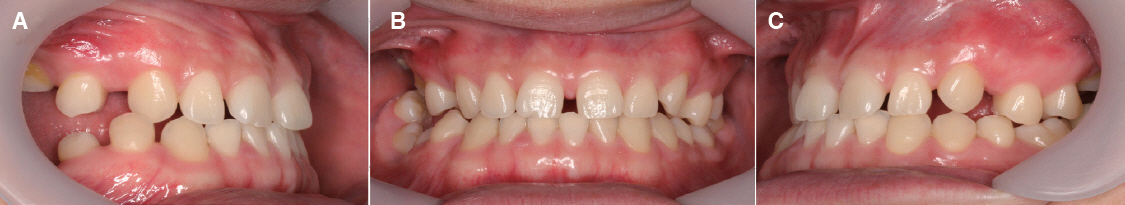
Fig. 1 Pre-treatment intraoral view. (A) Lateral view (right side), (B) Frontal view, (C) Lateral view (left side).
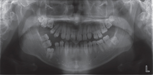
Fig. 2 Pre-treatment panoramic radiographic view.
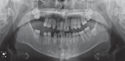
Fig. 3 Post-orthodontic treatment panoramic radiograpfic view.
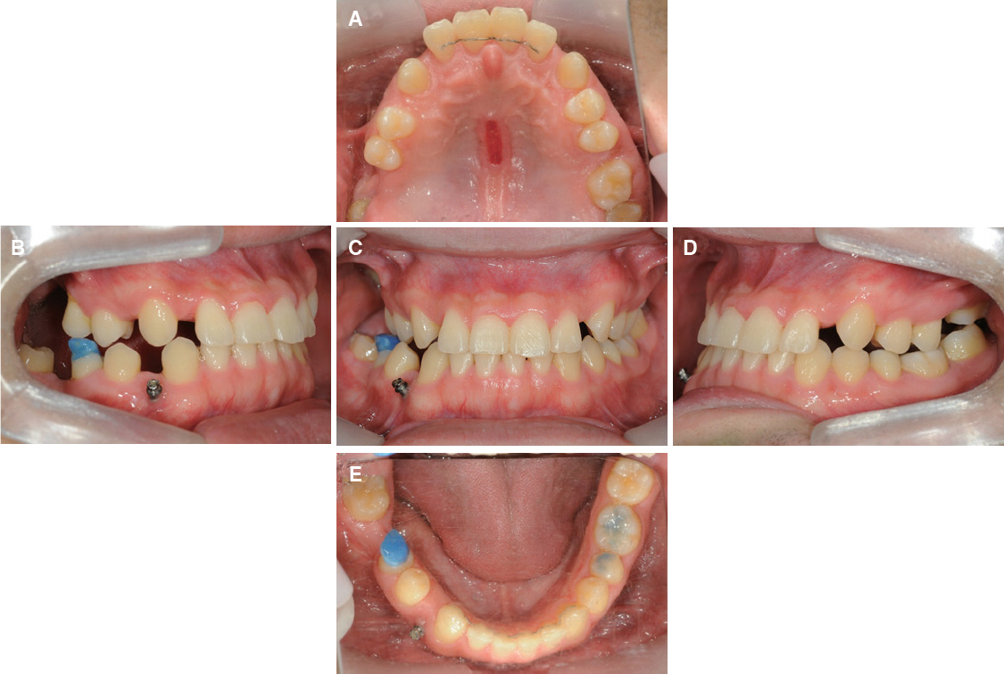
Fig. 4 Post-orthodontic treatment intraoral view. (A) Occlusal view of maxilla, (B) Lateral view (right side), (C) Frontal view, (D) Lateral view (left side), (E) Occlusal view of mandible.
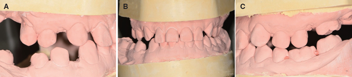
Fig. 5 Prognostic cast view. (A) Lateral view (right side), (B) Frontal view, (C) Lateral view (left side).
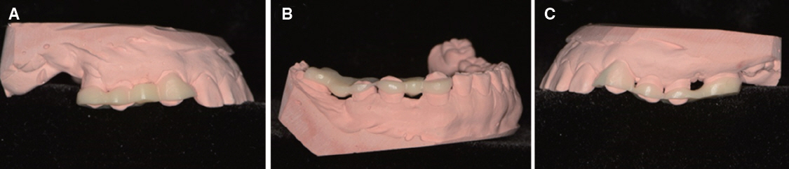
Fig. 6 Guide view. (A) Lateral view on Maxilla (right side), (B) Lateral view on Mandible (right side), (C) Lateral view on Maxilla (left side).
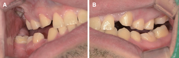
Fig. 7 Space view after prep tooth. (A) Lateral view (right side), (B) Lateral view (left side).
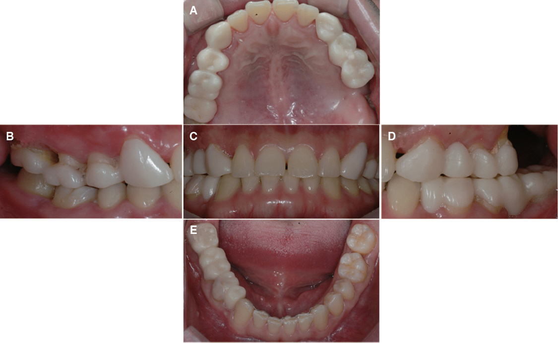
Fig. 8 Temporary crown setting intraoral view. (A) Occlusal view of maxilla, (B)Lateral view (right side), (C) Frontal view, (D) Lateral view (left side), (E) Occlusal view of mandible.
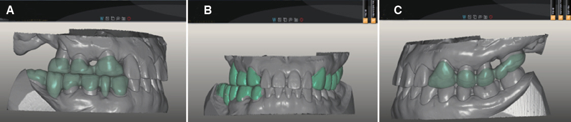
Fig. 9 Zirconia crown CAD view. (A) Lateral view (right side), (B) Frontal view, (C) Lateral view (left side).
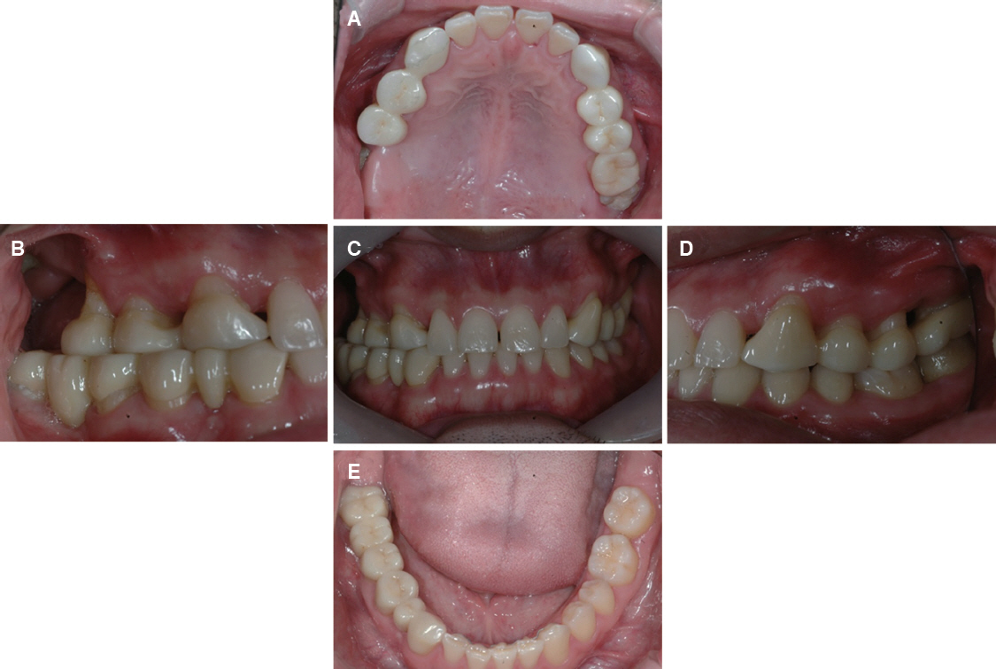
Fig. 10 Zirconia crown setting intraoral view. (A) Occlusal view of maxilla, (B) Lateral view (right side), (C) Frontal view, (D) Lateral view (left side), (E) Occlusal view of mandible.
Reference
-
References
1. Pahkala R, Pahkala A, Laine T. Eruption pattern of permanent teeth in a rural community in northeastern Finland. Acta Odontol Scand. 1991; 49:341–9. DOI: 10.3109/00016359109005930. PMID: 1776401.2. Nolla CM. The development of the human dentition. ASDC J Dent Child. 1960; 27:254–66.3. Kochhar R, Richardson A. The chronology and sequence of eruption of human permanent teeth in Northern Ireland. Int J Paediatr Dent. 1998; 8:24352. DOI: 10.1046/j.1365-263x.1998.00092.x.4. Peedikayil FC. Delayed tooth eruption. E-Journal of Dentistry. 2011; 1:81–6.5. Suri L, Gagari E, Vastardis H. Delayed tooth eruption:pathogenesis, diagnosis, and treatment. A literature review. Am J Orthod Dentofacial Orthop. 2004; 126:432–45. DOI: 10.1016/j.ajodo.2003.10.031. PMID: 15470346.6. Frazier-Bowers SA, Koehler KE, Ackerman JL, Proffit WR. Primary failure of eruption:further characterization of a rare eruption disorder. Am J Orthod Dentofacial Orthop. 2007; 131:578. DOI: 10.1016/j.ajodo.2006.09.038. PMID: 17482073.7. Kurol J. Early treatment of tooth-eruption disturbances. Am J Orthod Dentofacial Orthop. 2002; 121:588–91. DOI: 10.1067/mod.2002.124173. PMID: 12080309.8. Frank CA. Treatment options for impacted teeth. J Am Dent Assoc. 2000; 131:623–32. DOI: 10.14219/jada.archive.2000.0236. PMID: 10832256.9. Tsuo Y, Yoshida K, Atsuta M. Effects of aluminablasting and adhesive primers on bonding between resin luting agent and zirconia ceramics. Dent Mater J. 2006; 25:669–74. DOI: 10.4012/dmj.25.669. PMID: 17338299.10. The Fixed Prosthodontics Professor Association. 2012. 1st ed. Seoul: DaehanNarae Publishing Inc;p. 363–6.11. Hikita K, Van Meerbeek B, De Munck J, Ikeda T, Van Landuyt K, Maida T, Lambrechts P, Peumans M. Bonding effectiveness of adhesive luting agents to enamel and dentin. Dent Mater. 2007; 23:71–80. DOI: 10.1016/j.dental.2005.12.002. PMID: 16426673.12. Ritter AV, Ghaname E, Pimenta LA. Dentin and enamel bond strengths of dual-cure composite luting agents used with dual-cure dental adhesives. J Dent. 2009; 37:59–64. DOI: 10.1016/j.jdent.2008.09.006. PMID: 18926614.13. De Munck J, Vargas M, Van Landuyt K, Hikita K, Lambrechts P, Van Meerbeek B. Bonding of an auto-adhesive luting material to enamel and dentin. Dent Mater. 2004; 20:963–71. DOI: 10.1016/j.dental.2004.03.002. PMID: 15501325 .
