Primary Carcinosarcoma of the Fallopian Tube: A Case Report
- Affiliations
-
- 1Department of Radiology, Daegu Fatima Hospital, Daegu, Korea.
- 2Department of Radiology, Keimyung University School of Medicine, Dongsan Medical Center, Daegu, Korea. kseehdr@dsmc.or.kr
- 3Department of Pathology, Keimyung University School of Medicine, Dongsan Medical Center, Daegu, Korea.
- KMID: 2386743
- DOI: http://doi.org/10.3348/jksr.2017.77.2.85
Abstract
- Carcinosarcomas are biphasic neoplasms composed of epithelial and mesenchymal elements. They are most commonly found in the uterus, with rare involvement of the fallopian tubes. Here, we present a case of primary carcinosarcoma of the fallopian tube. On CT and MRI imaging, it manifested as a tubular heterogeneous enhancing mass, along with necrosis and hemorrhage.
Figure
-
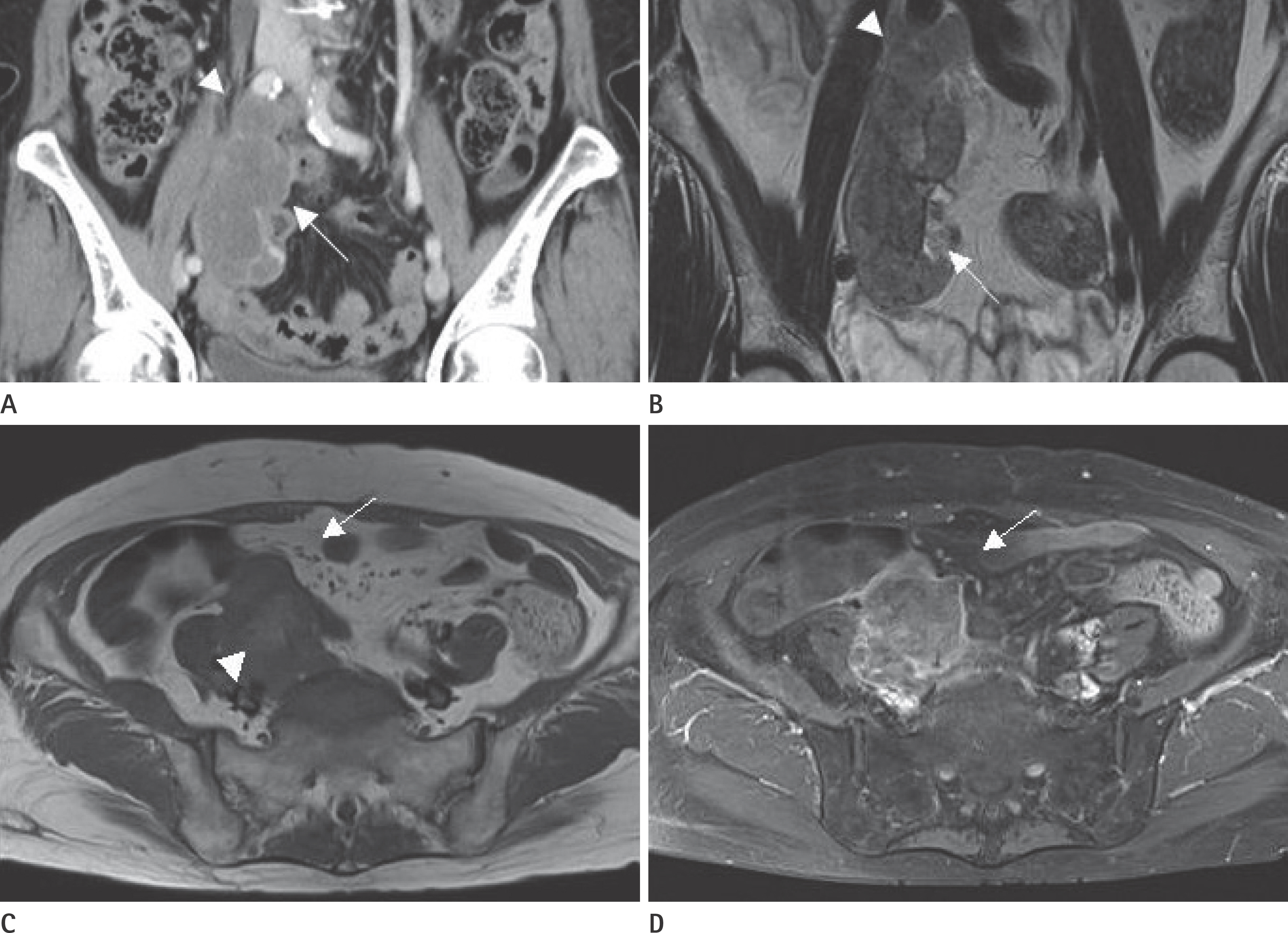
Fig. 1. 76-year-old female with primary fallopian carcinosarcoma. A. Coronal image of the dynamic abdominal CT scan demonstrates a lobulated hypoattenuating mass (arrow) and normal ovary (arrowhead). B. T2-weighted image shows a large heterogeneous, low signal intensity mass (arrow) and normal ovary (arrowhead). C. T1-weighted image shows relatively low signal intensity of tumor (arrow) with central regions of necrosis and hemorrhage in right fallopian tube (arrowhead). D. Contrast enhanced axial MRI shows heterogeneous enhancement of the tumor (arrow).
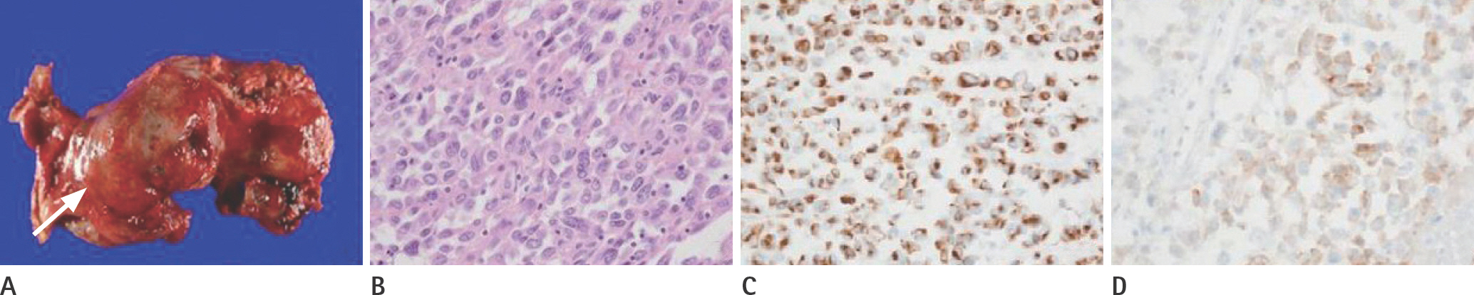
Fig. 2. Histopathologic findings of a surgical specimen in 76-year-old female with primary fallopian carcinosarcoma. A. Gross specimen: gross finding of right fallopian tube reveals markedly hemorrhagic and fusiform dilation with bulging tumor mass. The sero-sal surface is partially exposed by the tumor mass. However, right ovary is grossly intact (arrow). B. Microscopic examination (hematoxylin and eosin stain): high power field reveals spindle to round tumor cells with pleomorphic nuclei, vesicu-lar chromatin, and prominent nucleoli. C, D. Immunohistochemistry: diffuse strong positivity of vimentin (sarcoma component) (C) and focal positivity of epithelial membrane antigen; carcinoma component (× 400) (D).
Reference
-
1.Henderson SR., Harper RC., Salazar OM., Rudolph JH. Primary carcinoma of the fallopian tube: difficulties of diagnosis and treatment. Gynecol Oncol. 1977. 5:168–179.2.Mira A., Fereshteh F., Virginie F., Xavier SG. Primitive fallopian tube carcinosarcoma: three cases with immunohistochemi-cal profiling. Int J Cancer Clin Res. 2015. 1:2378–3419.
Article3.Hanjani P., Petersen RO., Bonnell SA. Malignant mixed mulle-rian tumor of the fallopian tube. Report of a case and re-view of literature. Gynecol Oncol. 1980. 9:381–393.4.Yang S., Lin L., Peng Z., Yang K., Lou J. Malignant mixed Mülle-rian tumor of the fallopian tube in a patient with irregular vaginal bleeding. Lab Med. 2009. 40:401–403.
Article5.Worthington JL., Balfe DM., Lee JK., Gersell DJ., Heiken JP., Ling D, et al. Uterine neoplasms: MR imaging. Radiology. 1986. 159:725–730.
Article6.Ohguri T., Aoki T., Watanabe H., Nakamura K., Nakata H., Mat-suura Y, et al. MRI findings including gadolinium-enhanced dynamic studies of malignant, mixed mesodermal tumors of the uterus: differentiation from endometrial carcinomas. Eur Radiol. 2002. 12:2737–2742.
Article7.Yamashita Y., Takahashi M., Miyazaki K., Okamura H. Contrast-enhanced MR imaging of malignant mixed müllerian tumor of the uterus. AJR Am J Roentgenol. 1993. 160:1150–1151.
Article8.Takemori M., Nishimura R., Yasuda D., Sugimura K. Carcino-sarcoma of the uterus: magnetic resonance imaging. Gy-necol Obstet Invest. 1997. 43:139–141.
Article9.Kawakami S., Togashi K., Kimura I., Nakano Y., Koshiyama M., Takakura K, et al. Primary malignant tumor of the fallopi-an tube: appearance at CT and MR imaging. Radiology. 1993. 186:503–508.
Article10.Lee HK., Kim SH., Cho JY., Yeon KM. Uterine adenofibroma and adenosarcoma: CT and MR findings. J Comput Assist Tomogr. 1998. 22:314–316.
