Thyroid Nodules with Macrocalcification: Sonographic Findings Predictive of Malignancy
- Affiliations
-
- 1Department of Radiology, Yonsei University College of Medicine, Gangnam Severance Hospital, Seoul, Korea. chrismd@hanmail.net
- 2Department of Radiology, Soonchunhyang University Hospital, Soonchunhyang University College of Medicine, Seoul, Korea.
- 3Department of Radiology and Research Institute of Radiological Science, Yonsei University College of Medicine, Severance Hospital, Seoul, Korea.
- 4Department of Surgery, Yonsei University College of Medicine, Gangnam Severance Hospital, Seoul, Korea.
Abstract
- PURPOSE
To analyze which sonographic features of thyroid nodules with macrocalcifications were predictable of thyroid malignancy.
MATERIALS AND METHODS
We reviewed sonographic findings of 854 macrocalcified thyroid nodules in patients who underwent fine needle aspiration biopsy between December 2009 and January 2011. There were 171 non-diagnostic aspirations, 34 nodules with category 3, 4, 5 based on Bethesda system, which were not confirmed by surgery, and these nodules were excluded from the analysis. Sonographic characteristics of the macrocalcifications including its thickness, interruption, and existence of soft tissue rim outside the macrocalcification were analyzed. Other sonographic characteristics of nodules such as shape, margin, composition, echo pattern, vascularity, and underlying parenchymal echogenicity were also evaluated. The correlation of sonographic features with cytopathologic results and the diagnostic performance of sonographic features for the prediction of malignancy were analyzed.
RESULTS
Among 649 nodules, 179 (27.6%) nodules were malignant and 470 (72.4%) nodules were benign. Among the features of the macrocalcification, interruption, irregular thickness, or the presence of soft tissue outside calcification rim were associated with malignancy (p<0.001). A high sensitivity and negative predictive values for the prediction of malignancy was found in sonographic characteristics of irregular thickness (92.2% and 91.0%, respectively) and the presence of soft tissue (88.5% and 88.8%, respectively).
CONCLUSION
Sonographic characteristics of macrocalcification such as interruption, irregular thickness and the presence of soft tissue rim were associated with malignancy in thyroid nodules with macrocalcifications.
MeSH Terms
Figure
-
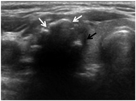
Fig. 1 US findings of malignant thyroid nodule with macrocalcification. Transverse US image in a 42-year-old woman shows a nodule with interrupted macrocalcification (white arrows), irregular thickness and soft tissue rim outside the calcification (black arrow) and the nodule was diagnosed as papillary thyroid carcinoma by fine needle aspiration cytology.
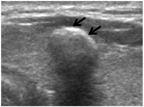
Fig. 2 US findings of benign thyroid nodule with macrocalcification. Transverse US image of a 54-year-old woman shows a nodule with macrocalcification (arrows) which has regular margin and regular thickness without interruption. The soft tissue rim outside of the macrocalcification was not visible and the nodule was diagnosed as adenomatous hyperplasia by fine needle aspiration biopsy.
Reference
-
1. Kim MJ, Kim EK, Kwak JY, Park CS, Chung WY, Nam KH, et al. Differentiation of thyroid nodules with macrocalcifications: role of suspicious sonographic findings. J Ultrasound Med. 2008; 27:1179–1184.2. Taki S, Terahata S, Yamashita R, Kinuya K, Nobata K, Kakuda K, et al. Thyroid calcifications: sonographic patterns and incidence of cancer. Clin Imaging. 2004; 28:368–371.3. Choi SH, Han KH, Yoon JH, Moon HJ, Son EJ, Youk JH, et al. Factors affecting inadequate sampling of ultrasound-guided fine-needle aspiration biopsy of thyroid nodules. Clin Endocrinol (Oxf). 2011; 74:776–782.
Article4. Alexander EK, Heering JP, Benson CB, Frates MC, Doubilet PM, Cibas ES, et al. Assessment of nondiagnostic ultrasound-guided fine needle aspirations of thyroid nodules. J Clin Endocrinol Metab. 2002; 87:4924–4927.
Article5. Yoon DY, Lee JW, Chang SK, Choi CS, Yun EJ, Seo YL, et al. Peripheral calcification in thyroid nodules: ultrasonographic features and prediction of malignancy. J Ultrasound Med. 2007; 26:1349–1355.6. Park M, Shin JH, Han BK, Ko EY, Hwang HS, Kang SS, et al. Sonography of thyroid nodules with peripheral calcifications. J Clin Ultrasound. 2009; 37:324–328.
Article7. Cibas ES, Ali SZ. NCI Thyroid FNA State of the Science Conference. The Bethesda System For Reporting Thyroid Cytopathology. Am J Clin Pathol. 2009; 132:658–665.
Article8. Carmeci C, Jeffrey RB, McDougall IR, Nowels KW, Weigel RJ. Ultrasound-guided fine-needle aspiration biopsy of thyroid masses. Thyroid. 1998; 8:283–289.
Article9. Danese D, Sciacchitano S, Farsetti A, Andreoli M, Pontecorvi A. Diagnostic accuracy of conventional versus sonography-guided fine-needle aspiration biopsy of thyroid nodules. Thyroid. 1998; 8:15–21.
Article10. Goellner JR, Gharib H, Grant CS, Johnson DA. Fine needle aspiration cytology of the thyroid, 1980 to 1986. Acta Cytol. 1987; 31:587–590.11. Rago T, Vitti P. Role of thyroid ultrasound in the diagnostic evaluation of thyroid nodules. Best Pract Res Clin Endocrinol Metab. 2008; 22:913–928.
Article12. Bastin S, Bolland MJ, Croxson MS. Role of ultrasound in the assessment of nodular thyroid disease. J Med Imaging Radiat Oncol. 2009; 53:177–187.
Article13. Seningen JL, Nassar A, Henry MR. Correlation of thyroid nodule fine-needle aspiration cytology with corresponding histology at Mayo Clinic, 2001-2007: an institutional experience of 1,945 cases. Diagn Cytopathol. 2012; 40:Suppl 1. E27–E32.
Article14. Renshaw AA. Histologic follow-up of nondiagnostic thyroid fine needle aspirations: implications for adequacy criteria. Diagn Cytopathol. 2012; 40:Suppl 1. E13–E15.
Article15. Berker D, Isik S, Ozuguz U, Tutuncu YA, Kucukler K, Akbaba G, et al. Prevalence of incidental thyroid cancer and its ultrasonographic features in subcentimeter thyroid nodules of patients with hyperthyroidism. Endocrine. 2011; 39:13–20.
Article16. Qian M, Wang J, Qiu Y. [The significance of calcification in the thyroid papillary carcinoma]. Lin Chung Er Bi Yan Hou Tou Jing Wai Ke Za Zhi. 2011; 25:673–675.17. Petrone L, Mannucci E, De Feo ML, Parenti G, Biagini C, Panconesi R, et al. A simple ultrasound score for the identification of candidates to fine needle aspiration of thyroid nodules. J Endocrinol Invest. 2012; 35:720–724.18. Ozel A, Erturk SM, Ercan A, Yılmaz B, Basak T, Cantisani V, et al. The diagnostic efficiency of ultrasound in characterization for thyroid nodules: how many criteria are required to predict malignancy? Med Ultrason. 2012; 14:24–28.19. Chen CY, Tseng HS, Lee CH, Chan WP. Primary squamous cell carcinoma of the thyroid gland with eggshell calcification: sonographic and computed tomographic findings. J Ultrasound Med. 2010; 29:1667–1670.
Article20. Yaturu S, Rainer L. Thyroid nodule with eggshell calcification and oncocytic thyroid cancer. Med Sci Monit. 2010; 16:CS25–CS28.21. Gooding GA. Ultrasonic appearance of a thyroid nodule invested in eggshell calcification. J Clin Ultrasound. 1978; 6:41–43.
Article22. Moon WJ, Jung SL, Lee JH, Na DG, Baek JH, Lee YH, et al. Benign and malignant thyroid nodules: US differentiation--multicenter retrospective study. Radiology. 2008; 247:762–770.
Article
- Full Text Links
-
- Actions
-
Cited
- CITED
-
- Close
- Share
-
- Similar articles
-
- Relationship of Shape of Macrocalcification and Thyroid Cancer: Correlation with US and Pathologic Findings
- Otolaryngologist-Performed Ultrasound and Ultrasound-Guided Fine Needle Aspiration for Thyroid Nodule and Meaningful Ultrasound Finding
- Malignancy Risk Stratification of Thyroid Nodules with Macrocalcification and Rim Calcification Based on Ultrasound Patterns
- Thyroid nodules with isolated macrocalcification: malignancy risk and diagnostic efficacy of fine-needle aspiration and core needle biopsy
- Sonographic Evaluation of Thyroid Nodules
