Normal Magnetic Resonance Perfusion Imaging and Atypical Posterior Reversible Encephalopathy Syndrome in Chronic Kidney Disease
- Affiliations
-
- 1Department of Neurology, Kangdong Sacred Heart Hospital, Hallym University College of Medicine, Seoul, Korea.
- 2Department of Neurology, Bucheon St. Mary's Hospital, College of Medicine, The Catholic University of Korea, Bucheon, Korea. ysshim@catholic.ac.kr
- KMID: 2385886
- DOI: http://doi.org/10.18700/jnc.170003
Abstract
- BACKGROUND
Posterior reversible encephalopathy syndrome (PRES) is classically characterized by symmetric vasogenic edema in the parietooccipital areas, but may occur at other sites with varying imaging appearances.
CASE REPORT
A 55-year old female with chronic kidney disease (CKD) was admitted to the emergency room, presenting with nausea, vomiting and seizure. The initial blood pressure was 145/90 mmHg. Fluid attenuated inversion recovery demonstrated diffuse vasogenic edema in the bilateral cortical and subcortical white matters involving the frontal lobes. Perfusion magnetic resonance imaging (MRP) showed no hyper- or hypoperfusion at blood pressure levels of 140/50 mmHg. A follow-up magnetic resonance imaging at 3 weeks later demonstrated complete resolution of previous lesions.
CONCLUSIONS
Earlier reports have demonstrated that PRES can occur in cases of atypical distributions, and features of imaging findings and normotensive settings. It is important to note that PRES is a dynamic process. As a result, we suggest that MRP must be considered in the appropriate temporal framework, to avoid misinterpretation of the other diseases, especially in CKD patients.
Keyword
MeSH Terms
Figure
-
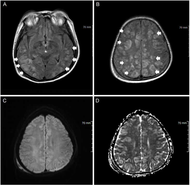
Figure 1. Initial brain-magnetic resonance imaging. (A, B) FLAIR demonstrated diffuse vasogenic edema in the bilateral cortical and subcortical white matters involving the frontal lobes (arrows). (C) DWI revealed non-significant high signal. (D) ADC map demonstrated high signal intensities in these regions. FLAIR, fluid attenuated inversion recovery; DWI, diffusion-weighted imaging; ADC, apparent diffusion coefficient.
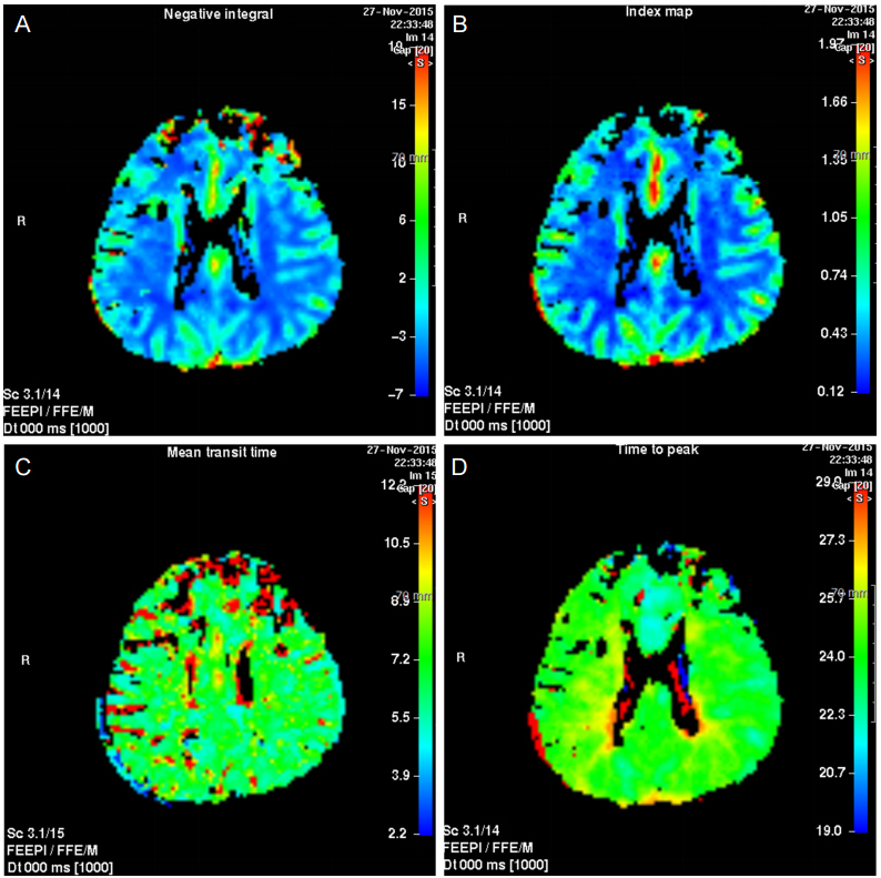
Figure 2. Normal perfusion MRI at blood pressure levels of 140/50 mmHg. (A) Cerebral blood volume, (B) cerebral blood flow, (C) mean transit time, (D) time to peak. MRI, magnetic resonance imaging.
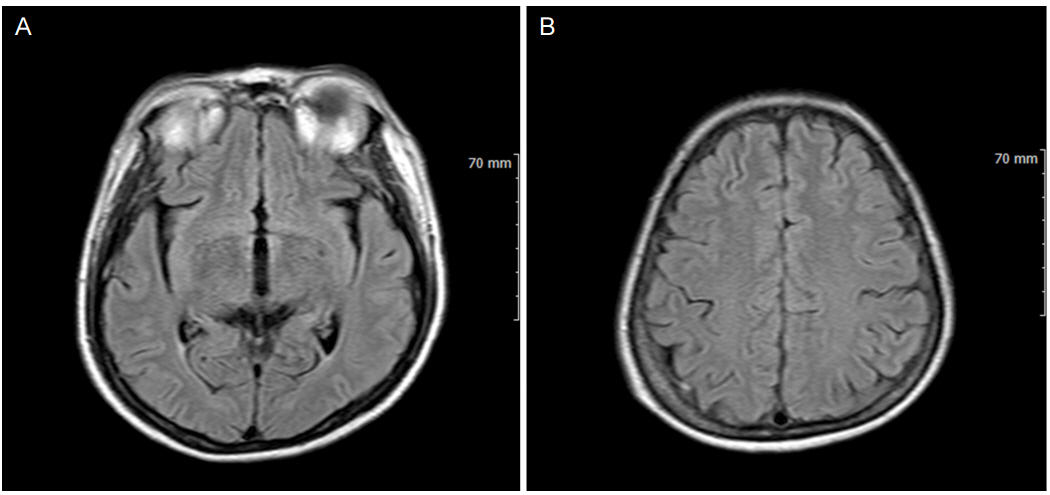
Figure 3. FLAIR after 3 weeks. The follow-up magnetic resonance imaging demonstrated complete resolution of previous lesions. FLAIR, fluid attenuated inversion recovery
Reference
-
1. Fugate JE, Claassen DO, Cloft HJ, Kallmes DF, Kozak OS, Rabinstein AA. Posterior reversible encephalopathy syndrome: associated clinical and radiologic findings. Mayo Clin Proc. 2010; 85:427–32.
Article2. Bartynski WS. Posterior reversible encephalopathy syndrome, part 1: fundamental imaging and clinical features. AJNR Am J Neuroradiol. 2008; 29:1036–42.
Article3. Hobson EV, Craven I, Blank SC. Posterior reversible encephalopathy syndrome: a truly treatable neurologic illness. Perit Dial Int. 2012; 32:590–4.
Article4. McKinney AM, Short J, Truwit CL, McKinney ZJ, Kozak OS, SantaCruz KS, et al. Posterior reversible encephalopathy syndrome: incidence of atypical regions of involvement and imaging findings. AJR Am J Roentgenol. 2007; 189:904–12.
Article5. Bartynski WS. Posterior reversible encephalopathy syndrome, part 2: controversies surrounding pathophysiology of vasogenic edema. AJNR Am J Neuroradiol. 2008; 29:1043–9.
Article6. Stevens CJ, Heran MK. The many faces of posterior reversible encephalopathy syndrome. Br J Radiol. 2012; 85:1566–75.
Article7. Bartynski WS, Boardman JF. Catheter angiography, MR angiography, and MR perfusion in posterior reversible encephalopathy syndrome. AJNR Am J Neuroradiol. 2008; 29:447–55.
Article8. Schwartz R, Mulkern R, Vajapeyam S, Kacher DF. Catheter angiography, MR angiography, and MR perfusion in posterior reversible encephalopathy syndrome. AJNR Am J Neuroradiol. 2009; 30:E19.
Article
- Full Text Links
-
- Actions
-
Cited
- CITED
-
- Close
- Share
-
- Similar articles
-
- A Case of Posterior Reversible Encephalopathy Syndrome in a Patient having Continuous Ambulatory Peritoneal Dialysis
- Hypertensive Brainstem Encephalopathy Combined with Acute Ischemic Stroke
- Posterior Reversible Encephalopathy Syndrome Following Meningitis in Pregnancy
- Posterior reversible encephalopathy syndrome caused by presumed Takayasu arteritis
- Hypertension-induced Posterior Reversible Encephalopathy Syndrome
