Progressive Occipitocervical Subluxation Despite Biologic Agent Therapy in a Patient with Advanced Rheumatoid Arthritis
- Affiliations
-
- 1Division of Rheumatology, Department of Internal Medicine, SMG-SNU Boramae Medical Center, Seoul, Korea. kideb1@gmail.com
- 2Department of Radiology, SMG-SNU Boramae Medical Center, Seoul National University College of Medicine, Seoul, Korea.
- KMID: 2385346
- DOI: http://doi.org/10.4078/jrd.2017.24.3.169
Abstract
- No abstract available.
MeSH Terms
Figure
-
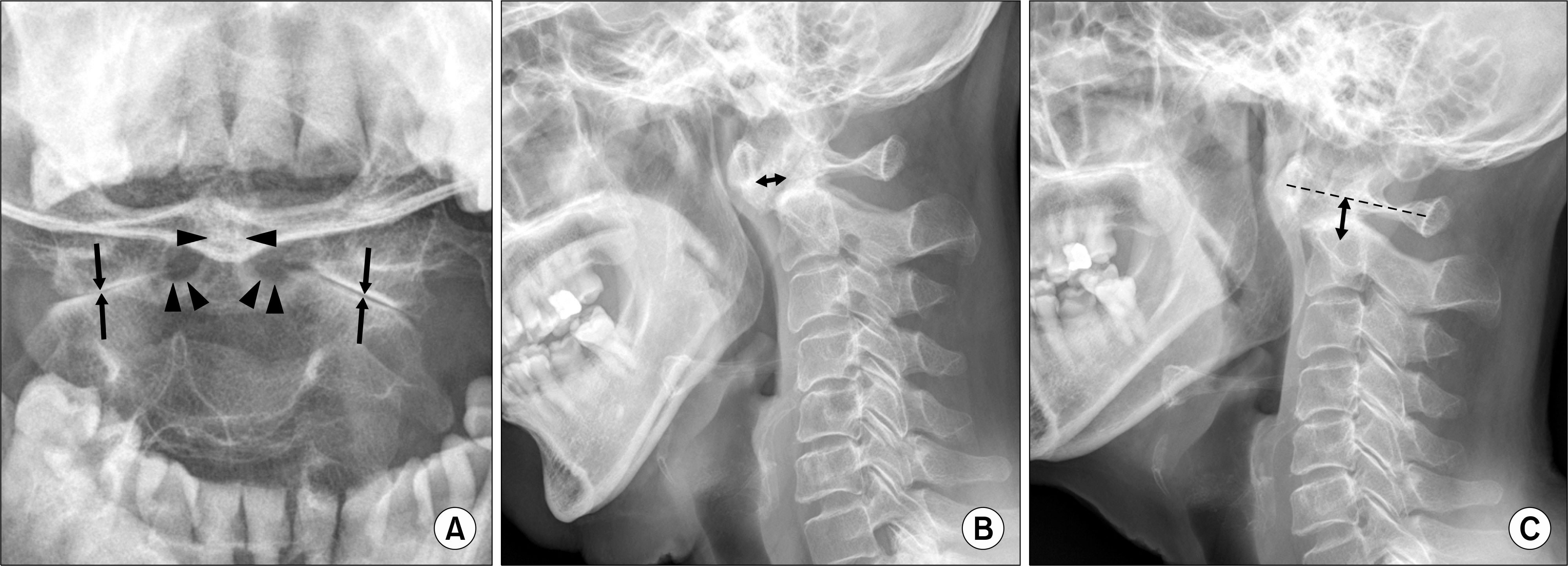
Figure 1. (A) There are multiple erosions (arrowheads) at the base and neck of the odontoid process of axis (open mouth view). Note the symmetrical joint space narrowing (arrows) of lateral atlantoaxial joints, which is a typical finding in rheumatoid arthritis.(B) Anterior subluxation of the atlantoaxial joint is evident at flexion. The atlantodental interval is 7 mm (double-headed arrow). (C) The Ranawat C1∼ C2 index (double-headed arrow) is 12 mm, just below the normal range.

Figure 2. (A) Three years later, the Ranawat C1∼ C2 index (double-headed arrow) is markedly decreased, suggesting basilar impression. Note the odontoid process (asterisk) is protruded beyond the horizontal axis of atlas (dashed line). (B) The reformatted sagittal computed tomography (CT) scan shows that the odontoid process is protruded above the level of foramen magnum (McRae line; dashed line). There are multiple erosions (arrows) in the C4, C5 discovertebral area. (C) A reformatted sagittal CT scan of a 25-year-old healthy female. The odontoid process is under the McRae line (dashed line).
Reference
-
1. Joaquim AF, Appenzeller S. Cervical spine involvement in rheumatoid arthritis–a systematic review. Autoimmun Rev. 2014; 13:1195–202.2. Ahn JK, Hwang JW, Oh JM, Lee J, Lee YS, Jeon CH, et al. Risk factors for development and progression of atlantoaxial subluxation in Korean patients with rheumatoid arthritis. Rheumatol Int. 2011; 31:1363–8.
Article3. Imagama S, Oishi Y, Miura Y, Kanayama Y, Ito Z, Wakao N, et al. Predictors of aggravation of cervical spine instability in rheumatoid arthritis patients: the large joint index. J Orthop Sci. 2010; 15:540–6.
Article4. Joaquim AF, Ghizoni E, Tedeschi H, Appenzeller S, Riew KD. Radiological evaluation of cervical spine involvement in rheumatoid arthritis. Neurosurg Focus. 2015; 38:E4.
Article5. Han MH, Ryu JI, Kim CH, Kim JM, Cheong JH, Bak KH, et al. Factors that predict risk of cervical instability in rheumatoid arthritis patients. Spine (Phila Pa. 1976; 2016 Oct 25 [Epub].DOI: DOI: 10.1097/BRS.0000000000001942.
Article
- Full Text Links
-
- Actions
-
Cited
- CITED
-
- Close
- Share
-
- Similar articles
-
- A Case of Rheumatoid Arthritis Presenting Initially as Atlantoaxial Subluxation
- Application of C1-C3 Halifax Interlaminar Clamps in Addition to C1-C2 Cable Fixation
- One-stage Transoral Decompression and Posterior Fusion in Rheumatoid Atlanto-axial Subluxation: Report of A Case
- Rheumatoid Atlantoaxial Subluxation: Report of a Case
- Irreducible Spacer Subluxation after Total Ankle Replacement Arthroplasty in a Patient with Rheumatoid Arthritis: A Case Report
