Imaging Findings of Primary Angiomyolipoma of the Pancreas: A Case Report
- Affiliations
-
- 1Department of Radiology, Korea Cancer Center Hospital, Korea Institute of Radiological and Medical Sciences, Seoul, Korea. lcf0666@hanmail.net
- KMID: 2384729
- DOI: http://doi.org/10.3348/jksr.2017.77.1.9
Abstract
- Angiomyolipoma (AML), a part of a family of mesenchymal tumors, is a common fat-containing solid neoplasm. Kidney and liver are the main sites of AML; rarely, primary pancreatic AML has also been reported. Here, we present a case of pathologically proven primary pancreatic AML in a middle-aged female patient, based on multidetector computed tomography scan, endoscopic ultrasound, positron emission tomography, and magnetic resonance imaging findings.
MeSH Terms
Figure
-
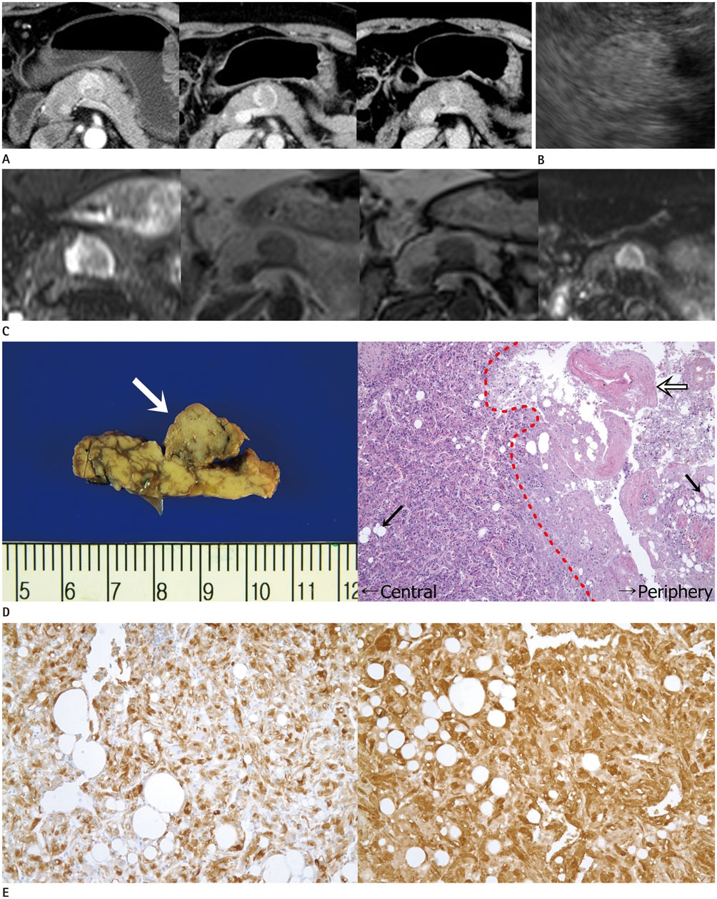
Fig. 1 A 59-year-old woman with primary angiomyolipoma of the pancreas. A. Axial contrast-enhanced MDCT scan series reveal a well-defined, 2-cm mass in the body of the pancreas without invasion of adjacent organs or vessels and no evidence of internal hemorrhage or necrosis. The mass demonstrates peripheral enhancement during the arterial (first image), pancreatic (second image), and portal venous phases (third image). B. EUS shows a hyperechogenic mass in the body of the pancreas, measuring 2-cm. C. MRI shows high peripheral and intermediate central SI on a HASTE T2-weighted image (first image). Both in-phase (second image) and opposed-phase (third image) images fail to demonstrate chemical shifting. A diffusion-weighted image (fourth image) reveals a high SI area in the pancreas without dark SI on the ADC map. This finding indicated the T2 shine-through artifact. ADC = apparent diffusion coefficient, EUS = endoscopic ultrasound, HASTE = half-Fourier acquisition single-shot turbo spin-echo, MDCT = multidetector computed tomography, SI = signal intensity D. Gross pathologic examination (left photograph) shows a 2 × 2 cm, well-defined, gray-white nodular tumor (white arrow) in the body of the pancreas. Hematoxylin and eosin staining (right photograph, ×100) reveals that the mass is composed of thick-walled blood vessels (open arrow), adipose tissue (black arrows), and spindle cells. Compared with the central area of the tumor, the peripheral area shows low cellularity with fibrinoid-like connective tissue. E. HMB-45 (left photograph) and SMA (right photograph) staining (immunohistochemistry, × 200) show diffusely positive immunoreactivity. Considering these results (D, E), the final pathologic diagnosis of the pancreatic tumor was determined as primary pancreatic AML. AML = angiomyolipoma, HMB-45 = human melanoma black-45, SMA = smooth muscle actin
Reference
-
1. Ferrozzi F, Zuccoli G, Bova D, Calculli L. Mesenchymal tumors of the pancreas: CT findings. J Comput Assist Tomogr. 2000; 24:622–627.2. Kim JY, Song JS, Park H, Byun JH, Song KB, Kim KP, et al. Primary mesenchymal tumors of the pancreas: single-center experience over 16 years. Pancreas. 2014; 43:959–968.3. Heywood G, Smyrk TC, Donohue JH. Primary angiomyolipoma of the pancreas. Pancreas. 2004; 28:443–445.4. Bhardwaj N, Garcea G, Lloyd D. A rare case of multi-focal angiomyolipoma affecting the pancreas and liver. J Surg Case Rep. 2012; 2012:5.5. Gleeson FC, de la Mora Levy JG, Zhang L, Levy MJ. The differential broadens. EUS FNA appearance and cytological findings of pancreatic angiomyolipoma. JOP. 2008; 9:67–70.6. Kim JK, Park SY, Shon JH, Cho KS. Angiomyolipoma with minimal fat: differentiation from renal cell carcinoma at biphasic helical CT. Radiology. 2004; 230:677–684.7. Israel GM, Hindman N, Hecht E, Krinsky G. The use of opposed-phase chemical shift MRI in the diagnosis of renal angiomyolipomas. AJR Am J Roentgenol. 2005; 184:1868–1872.8. Katabathina VS, Vikram R, Nagar AM, Tamboli P, Menias CO, Prasad SR. Mesenchymal neoplasms of the kidney in adults: imaging spectrum with radiologic-pathologic correlation. Radiographics. 2010; 30:1525–1540.9. Prasad SR, Wang H, Rosas H, Menias CO, Narra VR, Middleton WD, et al. Fat-containing lesions of the liver: radiologic-pathologic correlation. Radiographics. 2005; 25:321–331.10. Anderson SW, Kruskal JB, Kane RA. Benign hepatic tumors and iatrogenic pseudotumors. Radiographics. 2009; 29:211–229.
