Ann Dermatol.
2016 Oct;28(5):671-672. 10.5021/ad.2016.28.5.671.
Glandular Paget's Disease of the Male Nipple
- Affiliations
-
- 1Department of Dermatology, Inha University Hospital, Inha University School of Medicine, Incheon, Korea. jshin@inha.ac.kr
- KMID: 2382905
- DOI: http://doi.org/10.5021/ad.2016.28.5.671
Abstract
- No abstract available.
Figure
-
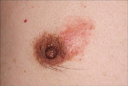
Fig. 1 A nonpruritic erythematous annular patch on the right areolar area.
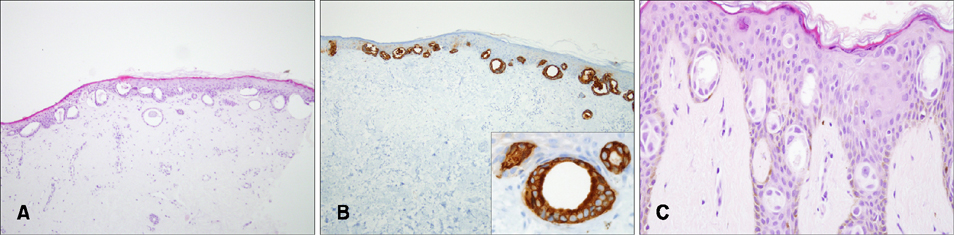
Fig. 2 (A) Punch biopsy revealed ductal structures with mild cellular atypia in the epidermis (H&E, ×100). (B) Immunohistochemistry for cytokeratin 7 was positive in the ductal cells (×40; inset, ×400). (C) The surgical specimen from Mohs micrographic surgery showed intraepidermal glandular structures, and Paget's cells—both single cells and nests (H&E, ×400).
Reference
-
1. Piñero A, Illana J, Martínez-Barba E, Sola J, Parrilla P. Extramammary Paget's disease of the breast: an unusual location with prognostic implications. Breast. 2005; 14:388–391.
Article2. Park YM, Kim JC, Choi JS, Kim KH. Paget's disease of the male breast. Ann Dermatol. 1992; 4:32–36.
Article3. Shousha S. Glandular Paget's disease of the nipple. Histopathology. 2007; 50:812–814.
Article4. Marucci G, Betts CM, Golouh R, Peterse JL, Foschini MP, Eusebi V. Toker cells are probably precursors of Paget cell carcinoma: a morphological and ultrastructural description. Virchows Arch. 2002; 441:117–123.
Article5. Toker C. Clear cells of the nipple epidermis. Cancer. 1970; 25:601–610.
Article
