Primary Glioblastoma of the Cerebellopontine Angle: Case Report and Review of the Literature
- Affiliations
-
- 1Department of Neurosurgery, VHS Medical Center, Seoul, Korea.
- 2Department of Neurosurgery, Korea University Guro Hospital, Korea University College of Medicine, Seoul, Korea. ns806@gmail.com
- KMID: 2382776
- DOI: http://doi.org/10.3340/jkns.2015.0303.006
Abstract
- Glioblastoma multiforme (GBM) is located most frequently in the cerebral hemispheres. Glioblastoma presenting as an extraaxial mass of cerebellopontine angle (CPA) is very rare in adults. We report a rare case of GBM arising in the CPA. The patient was a 71-year-old female, who complained of progressive gait disturbance and poor memory. Initial magnetic resonance imaging (MRI) revealed a 1.4×1.3 cm mass in the left CPA, with broad base to the petrous bone, showing homogenous enhancement. Follow-up MRI showed a rapid increase in size of mass (2.7×2.2 cm) with a necrotic portion. A stereotactic biopsy was done under the guidance of navigation system, and the histopathologic diagnosis was GBM, World Heath Organization grade IV. Further surgical resection was not performed considering her general condition, and the patient underwent concurrent chemotherapy with radiation therapy. Although rare, the possibility of glioblastoma should be included in the differential diagnosis of atypical CPA tumor.
MeSH Terms
Figure
-
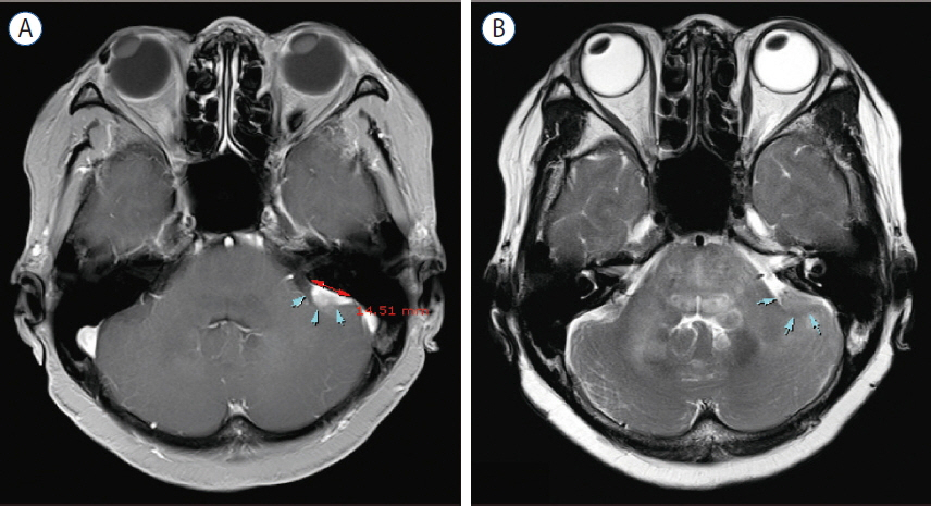
Fig. 1 Initial magnetic resonance imaging scans with axial enhanced T1-weighted (A) and T2-weighted (B) images, showing the mass in the left cere-bellopontine angle with broad base to the petrous bone (arrows).
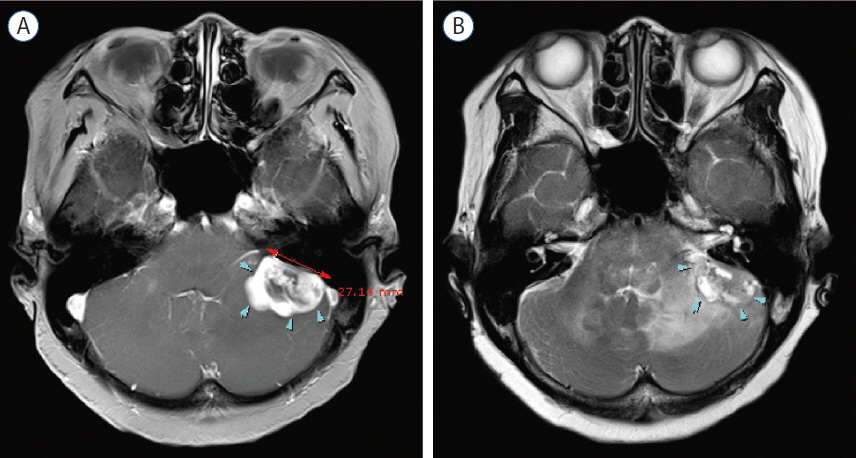
Fig. 2 3 months-follow-up magnetic resonance imaging scans with axial (A) T1-weighted and T2-weighted (B) images reveal rapid increase in size of mass and extensive peritumoral edema (arrows).
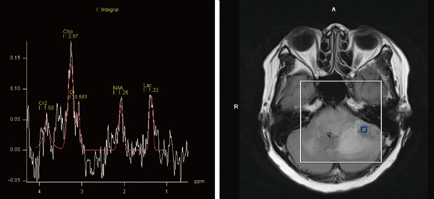
Fig. 3 Magnetic resonance spectroscopy demonstrates an increased choline to creatine ratio, decreased N-acetyl aspartate, and increased lactate. This findings indicates the possibility of high grade tumor.
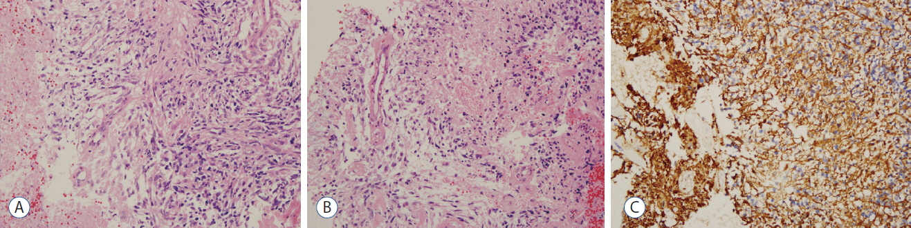
Fig. 4 A: Histopathology of the tumor shows atypical glial cells with frequent mitotic activity and moderate nuclear atypia (hematoxylin and eosin [H&E], 200×). B: Marked microvascular proliferation in the tumor (H&E, 200×). C: Immunohistochemical staining for GFAP reveals diffuse immunoreactivity in the tumor cells (200×). GFAP: glial fibrillary acidic protein.
Reference
-
References
1. Arnautovic KI, Husain MM, Linskey ME. Cranial nerve root entry zone primary cerebellopontine angle gliomas: a rare and poorly recognized subset of extraparenchymal tumors. J Neurooncol. 49:205–212. 2000.2. Kasliwal MK, Gupta DK, Mahapatra AK, Sharma MC. Multicentric cerebellopontine angle glioblastoma multiforme. Pediatr Neurosurg. 44:224–228. 2008.
Article3. Larjavaara S, Mäntylä R, Salminen T, Haapasalo H, Raitanen J, Jääskeläinen J, et al. Incidence of gliomas by anatomic location. Neuro Oncol. 9:319–325. 2007.
Article4. Linsenmann T, Monoranu CM, Westermaier T, Varallyay C, Ernestus RI, Vince GH. Exophytic glioblastoma arising from the cerebellum: case report and critical review of the literature. J Neurol Surg A Cent Eur Neurosurg. 74:262–264. 2013.
Article5. Matsuda M, Onuma K, Satomi K, Nakai K, Yamamoto T, Matsumura A. Exophytic cerebellar glioblastoma in the cerebellopontine angle: case report and review of the literature. J Neurol Surg Rep. 75:e67–e72. 2014.
Article6. Rasalingam K, Abdullah JM, Idris Z, Pal HK, Wahab N, Omar E, et al. A rare case of paediatric pontine glioblastoma presenting as a cerebellopontine angle otogenic abscess. Malays J Med Sci. 15:44–48. 2008.7. Salunke P, Sura S, Tewari MK, Gupta K, Khandelwal NK. An exophytic brain stem glioblastoma in an elderly presenting as a cerebellopontine angle syndrome. Br J Neurosurg. 26:96–98. 2012.
Article8. Stark AM, Maslehaty H, Hugo HH, Mahvash M, Mehdorn HM. Glioblastoma of the cerebellum and brainstem. J Clin Neurosci. 17:1248–1251. 2010.
Article9. Swaroop GR, Whittle IR. Exophytic pontine glioblastoma mimicking acoustic neuroma. J Neurosurg Sci. 41:409–411. 1997.10. Wu B, Liu W, Zhu H, Feng H, Liu J. Primary glioblastoma of the cerebellopontine angle in adults. J Neurosurg. 114:1288–1293. 2011.
Article11. Yamamoto M, Fukushima T, Sakamoto S, Tsugu H, Nagasaka S, Hirakawa K, et al. Cerebellar gliomas with exophytic growth--three case reports. Neurol Med Chir (Tokyo). 37:411–415. 1997.
- Full Text Links
-
- Actions
-
Cited
- CITED
-
- Close
- Share
-
- Similar articles
-
- Cerebellopontine Angle Medulloblastoma: Case Report
- Primary Intracranial Epidermoid Carcinoma
- Cystic Trigeminal Neurinoma at Cerebellopontine Angle
- Primary Choroid Plexus Papilloma of the Cerebellopontine Angle with Spinal Leptomeningeal Seeding
- A Case of Parasitic Cyst from Sparganosis in the Cerebellopontine Angle
