Surface changes of metal alloys and high-strength ceramics after ultrasonic scaling and intraoral polishing
- Affiliations
-
- 1Department of Prosthodontics, School of Dentistry, Seoul National University, Seoul, Republic of Korea.
- 2Department of Clinical Oral Health Science Graduate School of Clinical Dentistry, Ewha Womans University, Seoul, Republic of Korea.
- 3Department of Dentistry, School of Medicine, Ewha Womans University, Seoul, Republic of Korea. prosth@ewha.ac.kr
- KMID: 2382605
- DOI: http://doi.org/10.4047/jap.2017.9.3.188
Abstract
- PURPOSE
This study was to evaluate the effect of repeated ultrasonic scaling and surface polishing with intraoral polishing kits on the surface roughness of three different restorative materials.
MATERIALS AND METHODS
A total of 15 identical discs were fabricated with three different materials. The ultrasonic scaling was conducted for 20 seconds on the test surfaces. Subsequently, a multi-step polishing with recommended intraoral polishing kit was performed for 30 seconds. The 3D profiler and scanning electron microscopy were used to investigate surface integrity before scaling (pristine), after scaling, and after surface polishing for each material. Non-parametric Friedman and Wilcoxon signed rank sum tests were employed to statistically evaluate surface roughness changes of the pristine, scaled, and polished specimens. The level of significance was set at 0.05.
RESULTS
Surface roughness values before scaling (pristine), after scaling, and polishing of the metal alloys were 3.02±0.34 µm, 2.44±0.72 µm, and 3.49±0.72 µm, respectively. Surface roughness of lithium disilicate increased from 2.35±1.05 µm (pristine) to 28.54±9.64 µm (scaling), and further increased after polishing (56.66±9.12 µm, P<.05). The zirconia showed the most increase in roughness after scaling (from 1.65±0.42 µm to 101.37±18.75 µm), while its surface roughness decreased after polishing (29.57±18.86 µm, P<.05).
CONCLUSION
Ultrasonic scaling significantly changed the surface integrities of lithium disilicate and zirconia. Surface polishing with multi-step intraoral kit after repeated scaling was only effective for the zirconia, while it was not for lithium disilicate.
Figure
-
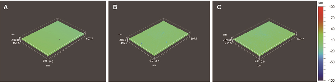
Fig. 1 A representative 3D plot image of metal (nickel-chromium) alloy; (A) pristine, (B) after repeated scaling for 20 seconds, (C) after surface polishing with an intraoral polishing kit.
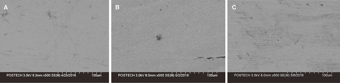
Fig. 2 A representative SEM image of metal (nickel-chromium) alloy (×500 magnification); (A) pristine, (B) after repeated scaling, (C) after surface polishing with an intraoral polishing kit.

Fig. 3 A representative 3D plot image of lithium disilicate; (A) pristine (glazed), (B) after repeated scaling for 20 seconds, (C) after multi-step polishing with an intraoral polishing kit.
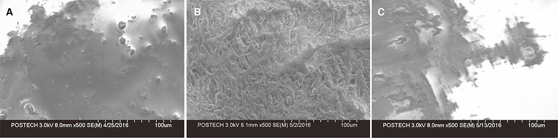
Fig. 4 A representative SEM image of lithium disilicate (×500 magnification); (A) pristine (glazed), (B) after repeated scaling, (C) after multi-step polishing with an intraoral polishing kit.
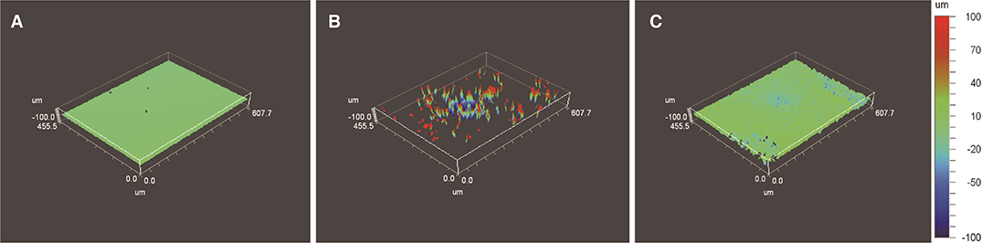
Fig. 5 A representative 3D plot image of zirconia; (A) pristine (glazed), (B) after repeated scaling for 20 seconds, (C) after multi-step polishing with an intraoral polishing kit.

Fig. 6 A representative SEM image of zirconia (×500 magnification); (A) pristine (glazed), (B) after repeated scaling, (C) after multi-step polishing with an intraoral polishing kit.
Reference
-
1. Axelsson P, Nyström B, Lindhe J. The long-term effect of a plaque control program on tooth mortality, caries and periodontal disease in adults. Results after 30 years of maintenance. J Clin Periodontol. 2004; 31:749–757.2. Axelsson P, Lindhe J. Effect of controlled oral hygiene procedures on caries and periodontal disease in adults. Results after 6 years. J Clin Periodontol. 1981; 8:239–248.3. Axelsson P, Lindhe J. The significance of maintenance care in the treatment of periodontal disease. J Clin Periodontol. 1981; 8:281–294.4. Pameijer CH, Stallard RE, Hiep N. Surface characteristics of teeth following periodontal instrumentation: a scanning electron microscope study. J Periodontol. 1972; 43:628–633.5. Breininger DR, O'Leary TJ, Blumenshine RV. Comparative effectiveness of ultrasonic and hand scaling for the removal of subgingival plaque and calculus. J Periodontol. 1987; 58:9–18.6. Sherman PR, Hutchens LH Jr, Jewson LG, Moriarty JM, Greco GW, McFall WT Jr. The effectiveness of subgingival scaling and root planning. I. Clinical detection of residual calculus. J Periodontol. 1990; 61:3–8.7. Drisko CH. Root instrumentation. Power-driven versus manual scalers, which one. Dent Clin North Am. 1998; 42:229–244.8. Kerry GJ. Roughness of root surfaces after use of ultrasonic instruments and hand curettes. J Periodontol. 1967; 38:340–346.9. Busslinger A, Lampe K, Beuchat M, Lehmann B. A comparative in vitro study of a magnetostrictive and a piezoelectric ultrasonic scaling instrument. J Clin Periodontol. 2001; 28:642–649.10. Oliveira G, Macedo PD, Tsurumaki JN, Sampaio JE, Marcantonio R. The effect of the angle of instrumentation of the Piezoelectric Ultrasonic Scaler on root surfaces. Int J Dent Hyg. 2016; 14:184–190.11. Wilkinson RF, Maybury JE. Scanning electron microscopy of the root surface following instrumentation. J Periodontol. 1973; 44:559–563.12. Kawai K, Urano M. Adherence of plaque components to different restorative materials. Oper Dent. 2001; 26:396–400.13. Quirynen M, Bollen CM. The influence of surface roughness and surface-free energy on supra- and subgingival plaque formation in man. A review of the literature. J Clin Periodontol. 1995; 22:1–14.14. Yilmaz C, Korkmaz T, Demirköprülü H, Ergün G, Ozkan Y. Color stability of glazed and polished dental porcelains. J Prosthodont. 2008; 17:20–24.15. Aykent F, Yondem I, Ozyesil AG, Gunal SK, Avunduk MC, Ozkan S. Effect of different finishing techniques for restorative materials on surface roughness and bacterial adhesion. J Prosthet Dent. 2010; 103:221–227.16. Weaks LM, Lescher NB, Barnes CM, Holroyd SV. Clinical evaluation of the Prophy-Jet as an instrument for routine removal of tooth stain and plaque. J Periodontol. 1984; 55:486–488.17. Lee SG, Lim SB, Chung CH, Kwon SH. Analysis of surface form change after performing prophylaxis procedure on implant surface using various oral hygiene instruments. J Korean Acad Periodontol. 2004; 34:1–17.18. Lee AR, Chung CH, Jung GU, Pang EK. The effect of copper alloy scaler tip on the surface roughness of dental implant and restorative materials. J Korean Acad Prosthodont. 2014; 52:177–185.19. Brecker SC. Porcelain baked to gold-A new medium in prosthodontics. J Prosthet Dent. 1956; 6:801–810.20. Della Bona A, Mecholsky JJ, Barrett AA, Griggs JA. Characterization of glass-infiltrated alumina-based ceramics. Dent Mater. 2008; 24:1568–1574.21. Kang JI, Heo YR, Lee MS, Son MK. Understanding and trends of esthetic treatment in prosthodontics: IPS e.max. J Korean Soc Dent Hyg. 2014; 14:447–452.22. Raigrodski AJ. Clinical and laboratory considerations for the use of CAD/CAM Y-TZP-based restorations. Pract Proced Aesthet Dent. 2003; 15:469–476.23. Conrad HJ, Seong WJ, Pesun IJ. Current ceramic materials and systems with clinical recommendations: a systematic review. J Prosthet Dent. 2007; 98:389–404.24. Kang JI, Heo YR, Lee MS, Son MK. Understanding and trends of esthetic treatment in prosthodontics: part 2. Zirconia. J Korean Soc Dent Hyg. 2014; 14:617–622.25. Volpato CAM, Fredel MC, Philippi AG, Petter CO. Ceramic materials and color in dentistry. INTECH Open Access Publisher;2010.26. Lee SC, Chung CH, Yim SB. The stereomicroscope and SPM study on the marginal change of porcelain crown in various repeated instrumentations for periodontal therapy. J Korean Acad Periodontol. 2000; 30:455–470.27. Vigolo P, Buzzo O, Buzzo M, Mutinelli S. An in vitro evaluation of alumina, zirconia, and lithium disilicate surface roughness caused by two scaling instruments. J Prosthodont. 2015; 12. 18.28. Patterson CJ, McLundie AC, Stirrups DR, Taylor WG. Efficacy of a porcelain refinishing system in restoring surface finish after grinding with fine and extra-fine diamond burs. J Prosthet Dent. 1992; 68:402–406.29. Ward MT, Tate WH, Powers JM. Surface roughness of opalescent porcelains after polishing. Oper Dent. 1995; 20:106–110.30. Chu FC, Frankel N, Smales RJ. Surface roughness and flexural strength of self-glazed, polished, and reglazed In-Ceram/Vitadur Alpha porcelain laminates. Int J Prosthodont. 2000; 13:66–71.31. Vrochari AD, Petropoulou A, Chronopoulos V, Polydorou O, Massey W, Hellwig E. Evaluation of surface roughness of ceramic and resin composite material used for conservative indirect restorations, after repolishing by intraoral means. J Prosthodont. 2015; 10. 21.32. Willems G, Lambrechts P, Braem M, Vuylsteke-Wauters M, Vanherle G. The surface roughness of enamel-to-enamel contact areas compared with the intrinsic roughness of dental resin composites. J Dent Res. 1991; 70:1299–1305.33. Haywood VB, Heymann HO, Scurria MS. Effects of water, speed, and experimental instrumentation on finishing and polishing porcelain intra-orally. Dent Mater. 1989; 5:185–188.34. Odatsu T, Jimbo R, Wennerberg A, Watanabe I, Sawase T. Effect of polishing and finishing procedures on the surface integrity of restorative ceramics. Am J Dent. 2013; 26:51–55.35. Sasahara RM, Ribeiro Fda C, Cesar PF, Yoshimura HN. Influence of the finishing technique on surface roughness of dental porcelains with different microstructures. Oper Dent. 2006; 31:577–583.36. Amaya-Pajares SP, Ritter AV, Vera Resendiz C, Henson BR, Culp L, Donovan TE. Effect of Finishing and Polishing on the Surface Roughness of Four Ceramic Materials after Occlusal Adjustment. J Esthet Restor Dent. 2016; 28:382–396.37. Preis V, Grumser K, Schneider-Feyrer S, Behr M, Rosentritt M. The effectiveness of polishing kits: influence on surface roughness of zirconia. Int J Prosthodont. 2015; 28:149–151.
- Full Text Links
-
- Actions
-
Cited
- CITED
-
- Close
- Share
-
- Similar articles
-
- Effect of Degassing Condition on Ceramic Bond Strength of Ni-Cr Alloys
- The effect of surface finishes on flexural strength, fracture toughness of feldspathic dental porcelain
- THE EFFECTS OF METAL SURFACE TREATMENTS ON THE BONE STRENGTH OF POLYMETHYL METHACRYLATE BONDED REMOVABLE PROSTHESE
- A study on surface roughness of metals according to finishing and polishing procedures: an atomic force microscope analysis
- Bond and fracture strength of metal-ceramic restorations formed by selective laser sintering
