A Case of Ophthalmic Artery Occlusion Following Subcutaneous Injection of Epinephrine Mixed with Lidocaine into the Supratrochlear Area
- Affiliations
-
- 1Retina Center, Department of Ophthalmology, HanGil Eye Hospital, Incheon, Korea.
- 2Department of Ophthalmology, Asan Medical Center, University of Ulsan College of Medicine, Seoul, Korea. junekim@amc.seoul.kr
- KMID: 2379886
- DOI: http://doi.org/10.3341/kjo.2017.0004
Abstract
- No abstract available.
Figure
-
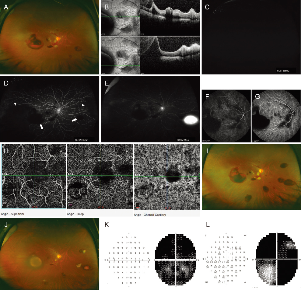
Fig. 1 (A) Wide fundus photograph of the right eye, 3 days after the initial presentation. Multiple cotton wool patches (CWPs) and retinal hemorrhages were observed around the optic nerve and macula. Large preretinal hemorrhages were observed at the nasal and temporal mid-peripheral retina. (B) Optical coherence tomography of the right eye, 3 days after the initial presentation. Preretinal hemorrhages of various sizes, posterior shadowing, and increased inner retinal hyperreflectivity (suggestive of ischemia of the inner retina) were observed. (C-E) Wide fluorescein angiography of the right eye, 3 days after the initial presentation. (C) A chorioretinal artery filling delay was seen in the early phase. (D) Multiple blocked fluorescent areas due to preretinal hemorrhages (arrows), and peripheral nonperfusion areas in the nasal and temporal retina (arrowheads) were noted in the arteriovenous phase. (E) Multiple vascular leakages and stains were observed in the late phase. (F,G) Indocyanine green angiography, 7 days after the initial presentation, revealed delayed filling of the choroidal artery in the early phase, and a segmented filling defect of the choriocapillaris. (H) Optical coherence tomography angiography (3 × 3 mm) exhibited disruption of the foveal avascular zone in the superficial and deep capillary plexuses, and multiple-capillary nonperfusion at the level of the superficial and deep retinal capillaries and choriocapillaris. (I) Immediately after the 3-day course of high-dose intravenous steroid therapy, wide fundus photography exhibited increased CWP sizes, retinal hemorrhages, and newly developed nasal and inferior retinal hemorrhages, compared with fundus findings at the first visit (A). (J) Four weeks after high-dose steroid treatment, most retinal hemorrhages and CWPs had disappeared. (K) Right eye 30-2 Humphrey visual field examination of the right eye, 3 days after initial presentation displaying extensive central, ceco-central and para-central scotomas. (L) Four weeks after high-dose steroid treatment, the visual field examination results revealed further deterioration; stimulus III could not be performed, and stimulus V displayed marked deterioration of the superonasal and temporal scotoma with inferonasal field sparing.
Reference
-
1. Niemi G. Advantages and disadvantages of adrenaline in regional anaesthesia. Best Pract Res Clin Anaesthesiol. 2005; 19:229–245.2. Park KH, Kim YK, Woo SJ, et al. Iatrogenic occlusion of the ophthalmic artery after cosmetic facial filler injections: a national survey by the Korean Retina Society. JAMA Ophthalmol. 2014; 132:714–723.3. Lazzeri D, Agostini T, Figus M, et al. Blindness following cosmetic injections of the face. Plast Reconstr Surg. 2012; 129:995–1012.4. Savino PJ, Burde RM, Mills RP. Visual loss following intranasal anesthetic injection. J Clin Neuroophthalmol. 1990; 10:140–144.5. Webber B, Orlansky H, Lipton C, Stevens M. Complications of an intra-arterial injection from an inferior alveolar nerve block. J Am Dent Assoc. 2001; 132:1702–1704.
- Full Text Links
-
- Actions
-
Cited
- CITED
-
- Close
- Share
-
- Similar articles
-
- Teh Effect of Local anesthetic on Degeneration and Regeneration of the Experimentally Traumatized Striated Muscle
- A Case of Unilateral Blindness Following Subcutaneous Injection of the silicone Oil on the Glabellar Area
- The Antimicrobial effects of Lidocaine and Epinephrine
- Effects of Epinephrine on Blood Concentration of Lidocaine and Epidural Block during Cesarean Section
- Ophthalmic Artery Occlusion After Carotid Revascularization
