The Effects of Multi-Growth Factors-Containing Cream on Post-Thyroidectomy Scars: A Preliminary Study
- Affiliations
-
- 1Department of Dermatology, Severance Hospital, Seoul, Korea. juhee@yuhs.ac
- 2Cutaneous Biology Research Institute, Yonsei University College of Medicine, Seoul, Korea.
- 3Department of Surgery, Yonsei University College of Medicine, Seoul, Korea.
- 4Institute of Endocrine Research, Yonsei University College of Medicine, Seoul, Korea.
- KMID: 2378531
- DOI: http://doi.org/10.5021/ad.2017.29.3.314
Abstract
- BACKGROUND
Growth factors play important roles in wound healing. However, the evidence for the effects of growth factors on post-thyroidectomy scars is limited.
OBJECTIVE
We performed a prospective study to assess the preventive and therapeutic effect of a multi-growth factor (MGF)-containing cream on post-thyroidectomy scars.
METHODS
Twenty-one patients with thyroidectomy scars applied MGF cream twice a day. We assessed the changes in erythema, pigmentation, skin elasticity, and skin hydration status using the erythema index, melanin index, cutometer, and corneometer, respectively. In addition, Vancouver scar scale (VSS) and patient satisfaction were assessed at 10 days after surgery (baseline), 2 weeks, 6 weeks, and 12 weeks after baseline.
RESULTS
The mean total VSS scores were significantly lower at 6 weeks (3.24±1.51 vs. 1.91±1.38) and 12 weeks (3.24±1.51 vs. 1.71±1.59) compared to the baseline. The degree of pigmentation was significantly lower at 12 weeks compared to the baseline, and the skin elasticity, and the skin hydration status were significantly higher at 12 weeks compared to the baseline. Over 85% of the patients were satisfied with the use of MGF cream without any adverse effect.
CONCLUSION
MGF cream might have additive or supportive effect for scar formation after thyroidectomy.
Keyword
MeSH Terms
Figure
-
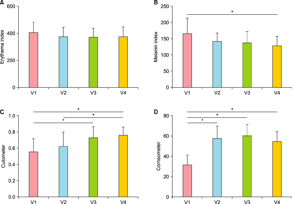
Fig. 1 (A~D) Data from the objective assessments of the patients at every visit. V1: visit 1 (baseline), V2: visit 2 (2 weeks after applying multi-growth factor [MGF] cream), V3: visit 3 (6 weeks after applying MGF cream), V4: visit 4 (12 weeks after applying MGF cream). *Signifies statistically significant difference (p<0.05).

Fig. 2 (A~E) Vancouver scar scale (VSS) score of the patients at every visit. V1: visit 1 (baseline), V2: visit 2 (2 weeks after applying multi-growth factor [MGF] cream), V3: visit 3 (6 weeks after applying MGF cream), V4: visit 4 (12 weeks after applying MGF cream). *Signifies statistically significant difference (p <0.05).

Fig. 3 Photographs of a 48-year-old male patient taken at each visit. (A) Baseline (Vancouver scar scale [VSS] vascularity 1, pigmentation 1, pliability 1, height 0), (B) 2 weeks after applying multi-growth factor (MGF) cream (VSS vascularity 0, pigmentation 0, pliability 1, height 0), (C) 6 weeks after applying MGF cream (VSS vascularity 0, pigmentation 0, pliability 0, height 0), (D) 12 weeks after applying MGF cream (VSS vascularity 0, pigmentation 0, pliability 0, height 0). The post-thyroidectomy scar showed improvement after applying MGF cream.

Fig. 4 Overall satisfaction score after applying multi-growth factor cream.
Reference
-
1. Ahn HS, Kim HJ, Welch HG. Korea's thyroid-cancer “epidemic”--screening and overdiagnosis. N Engl J Med. 2014; 371:1765–1767.
Article2. Rahib L, Smith BD, Aizenberg R, Rosenzweig AB, Fleshman JM, Matrisian LM. Projecting cancer incidence and deaths to 2030: the unexpected burden of thyroid, liver, and pancreas cancers in the United States. Cancer Res. 2014; 74:2913–2921.
Article3. American Thyroid Association (ATA) Guidelines Taskforce on Thyroid Nodules and Differentiated Thyroid Cancer. Cooper DS, Doherty GM, Haugen BR, Kloos RT, Lee SL, et al. Revised American Thyroid Association management guidelines for patients with thyroid nodules and differentiated thyroid cancer. Thyroid. 2009; 19:1167–1214.
Article4. Fonder MA, Lazarus GS, Cowan DA, Aronson-Cook B, Kohli AR, Mamelak AJ. Treating the chronic wound: a practical approach to the care of nonhealing wounds and wound care dressings. J Am Acad Dermatol. 2008; 58:185–206.
Article5. Sullivan T, Smith J, Kermode J, McIver E, Courtemanche DJ. Rating the burn scar. J Burn Care Rehabil. 1990; 11:256–260.
Article6. Tuan TL, Nichter LS. The molecular basis of keloid and hypertrophic scar formation. Mol Med Today. 1998; 4:19–24.
Article7. Lumenta DB, Siepmann E, Kamolz LP. Internet-based survey on current practice for evaluation, prevention, and treatment of scars, hypertrophic scars, and keloids. Wound Repair Regen. 2014; 22:483–491.
Article8. Berman B, Bieley HC. Adjunct therapies to surgical management of keloids. Dermatol Surg. 1996; 22:126–130.
Article9. Ledon JA, Savas J, Franca K, Chacon A, Nouri K. Intralesional treatment for keloids and hypertrophic scars: a review. Dermatol Surg. 2013; 39:1745–1757.
Article10. Guo S, Dipietro LA. Factors affecting wound healing. J Dent Res. 2010; 89:219–229.
Article11. Diegelmann RF, Evans MC. Wound healing: an overview of acute, fibrotic and delayed healing. Front Biosci. 2004; 9:283–289.
Article12. Broughton G 2nd, Janis JE, Attinger CE. The basic science of wound healing. Plast Reconstr Surg. 2006; 117:7 Suppl. 12S–34S.
Article13. Velnar T, Bailey T, Smrkolj V. The wound healing process: an overview of the cellular and molecular mechanisms. J Int Med Res. 2009; 37:1528–1542.
Article14. Dejana E, Languino LR, Polentarutti N, Balconi G, Ryckewaert JJ, Larrieu MJ, et al. Interaction between fibrinogen and cultured endothelial cells. Induction of migration and specific binding. J Clin Invest. 1985; 75:11–18.
Article15. Grotendorst GR, Soma Y, Takehara K, Charette M. EGF and TGF-alpha are potent chemoattractants for endothelial cells and EGF-like peptides are present at sites of tissue regeneration. J Cell Physiol. 1989; 139:617–623.
Article16. Terranova VP, DiFlorio R, Lyall RM, Hic S, Friesel R, Maciag T. Human endothelial cells are chemotactic to endothelial cell growth factor and heparin. J Cell Biol. 1985; 101:2330–2334.
Article17. Nanney LB. Epidermal and dermal effects of epidermal growth factor during wound repair. J Invest Dermatol. 1990; 94:624–629.
Article18. Brown GL, Curtsinger L 3rd, Brightwell JR, Ackerman DM, Tobin GR, Polk HC Jr, et al. Enhancement of epidermal regeneration by biosynthetic epidermal growth factor. J Exp Med. 1986; 163:1319–1324.
Article19. Epstein JB, Gorsky M, Guglietta A, Le N, Sonis ST. The correlation between epidermal growth factor levels in saliva and the severity of oral mucositis during oropharyngeal radiation therapy. Cancer. 2000; 89:2258–2265.
Article20. Kim YS, Lew DH, Tark KC, Rah DK, Hong JP. Effect of recombinant human epidermal growth factor against cutaneous scar formation in murine full-thickness wound healing. J Korean Med Sci. 2010; 25:589–596.
Article21. Steed DL. Clinical evaluation of recombinant human platelet-derived growth factor for the treatment of lower extremity diabetic ulcers. Diabetic Ulcer Study Group. J Vasc Surg. 1995; 21:71–78. discussion 79-81.22. Steed DL. Clinical evaluation of recombinant human platelet-derived growth factor for the treatment of lower extremity ulcers. Plast Reconstr Surg. 2006; 117:7 Suppl. 143S–149S. discussion 150S-151S.
Article23. O'Brien L, Pandit A. Silicon gel sheeting for preventing and treating hypertrophic and keloid scars. Cochrane Database Syst Rev. 2006; (1):CD003826.24. Meaume S, Le Pillouer-Prost A, Richert B, Roseeuw D, Vadoud J. Management of scars: updated practical guidelines and use of silicones. Eur J Dermatol. 2014; 24:435–443.
Article25. Hanasono MM, Lum J, Carroll LA, Mikulec AA, Koch RJ. The effect of silicone gel on basic fibroblast growth factor levels in fibroblast cell culture. Arch Facial Plast Surg. 2004; 6:88–93.
Article26. Choi J, Lee EH, Park SW, Chang H. Regulation of transforming growth factor β1, platelet-derived growth factor, and basic fibroblast growth factor by silicone gel sheeting in early-stage scarring. Arch Plast Surg. 2015; 42:20–27.
Article
- Full Text Links
-
- Actions
-
Cited
- CITED
-
- Close
- Share
-
- Similar articles
-
- Impact of Postthyroidectomy Scar on the Quality of Life of Thyroid Cancer Patients
- Clinical Study of Post Thyroidectomy Hypocalcemia
- Effect of Relaxin Expressing Adenovirus on Scar Remodeling: A Preliminary Study
- Completion Thyroidectomy
- Assessment of Postoperative Scar Using Modified Vancouver Scar Scale of 283 Patients Who Underwent Open Thyroidectomy in a Single Institution
