Korean J Neurotrauma.
2015 Oct;11(2):106-111. 10.13004/kjnt.2015.11.2.106.
Clinical Experience of Infantile Posthemorrhagic Hydrocephalus Treated with Ventriculo-Peritoneal Shunt
- Affiliations
-
- 1Department of Neurosurgery, Daegu Catholic University College of Medicine, Daegu, Korea. gneuros@cu.ac.kr
- KMID: 2378267
- DOI: http://doi.org/10.13004/kjnt.2015.11.2.106
Abstract
OBJECTIVE
Infantile posthemorrhagic hydrocephalus (IPHH) is the most common cause of infantile acquired hydrocephalus. We present and discuss our experience of treatment of six IPHH patients treated by a ventriculo-peritoneal (VP) shunt.
METHODS
Six preterm infants treated by a VP shunt due to germinal matrix hemorrhage and hydrocephalus were included in our study. External ventricular drainage (EVD) was performed in patients with symptomatic ventricular dilatation, and a VP shunt was placed in the case of no improvement of the ventricular index despite several rounds of EVD. Radiographic findings and surgical outcomes were analyzed retrospectively.
RESULTS
Four patients were male and two were female. Mean gestational age was 25 weeks and mean weight at birth was 868.3 g. One patient had a Papile grade II (16.7%) hemorrhage, three had a grade III (50%) hemorrhage, and two had a grade IV (33.3%) hemorrhage. EVD complications (one case of ventriculitis and one case of a ventricular abscess) occurred in two patients. VP shunt complications occurred in two patients (33.3%). Three cases had an isolated 4th ventricle; two of these cases had a VP shunt placed whereas the other case had a VP shunt placed in addition to aqueductoplasty using a neuroendoscope. At the last follow-up, three of the six patients had severe neurodevelopmental delay, two had mild neurodevelopmental delay, and one had normal development status.
CONCLUSION
In our study, although it is difficult to present the significant result for management of IPHH, we think that varied efforts are required to treat IPHH patients.
Keyword
MeSH Terms
Figure
-
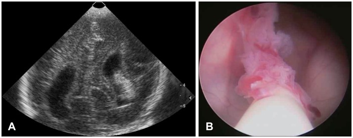
FIGURE 1 A case with complicated by proximal catheter occlusion. A: Ultrasonography taken seven days after birth shows intraventricular hemorrhage with dilatation of the lateral ventricle. B: Intraoperative neuroendoscopic view at the time of shunt revision shows encasement of choroid plexus around the intracranial catheter.
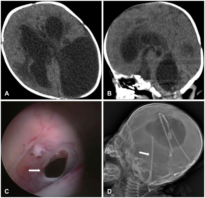
FIGURE 2 A case complicated by multiloculated ventricle. Multiple septations of lateral, 3rd, and 4th ventricle is shown in computed tomography (A, B). Sylvius aqueduct was reopened by a neuroendoscope (white arrow in C), and stent insertion into the Sylvius aqueduct is shown on the skull X-ray (white arrow in D).
Reference
-
1. Ballabh P. Intraventricular hemorrhage in premature infants: mechanism of disease. Pediatr Res. 2010; 67:1–8. PMID: 19816235.
Article2. Brouwer MJ, de Vries LS, Pistorius L, Rademaker KJ, Groenendaal F, Benders MJ. Ultrasound measurements of the lateral ventricles in neonates: why, how and when? A systematic review. Acta Paediatr. 2010; 99:1298–1306. PMID: 20394588.
Article3. Cinalli G, Spennato P, Savarese L, Ruggiero C, Aliberti F, Cuomo L, et al. Endoscopic aqueductoplasty and placement of a stent in the cerebral aqueduct in the management of isolated fourth ventricle in children. J Neurosurg. 2006; 104(1 Suppl):21–27. PMID: 16509476.
Article4. Cornips E, Van Calenbergh F, Plets C, Devlieger H, Casaer P. Use of external drainage for posthemorrhagic hydrocephalus in very low birth weight premature infants. Childs Nerv Syst. 1997; 13:369–374. PMID: 9298272.
Article5. Duncan CC, Ment LR. Intraventricular hemorrhage and prematurity. Neurosurg Clin N Am. 1993; 4:727–734. PMID: 8241793.
Article6. Fletcher JM, Landry SH, Bohan TP, Davidson KC, Brookshire BL, Lachar D, et al. Effects of intraventricular hemorrhage and hydrocephalus on the long-term neurobehavioral development of preterm very-low-birthweight infants. Dev Med Child Neurol. 1997; 39:596–606. PMID: 9344052.
Article7. Folkerth RD. Germinal matrix haemorrhage: destroying the brain's building blocks. Brain. 2011; 134(Pt 5):1261–1263. PMID: 21596767.
Article8. Hambleton G, Wigglesworth JS. Origin of intraventricular haemorrhage in the preterm infant. Arch Dis Child. 1976; 51:651–659. PMID: 999324.
Article9. Januschek E, Machado LS, Steinthal B, Ulrich PT. Posthemorrhagic hydrocephalus in very low birth weight infants--a new gentle surgical technique for external ventricular drainage. Childs Nerv Syst. 2011; 27:991–994. PMID: 21399966.
Article10. Kedzia A. Characteristics of periventricular matrix vascularization in image computer transformation system. Folia Neuropathol. 1995; 33:267–270. PMID: 8673438.11. Kim SY, Cho JH, Kim KH. Endoscopic coagulation of choroid plexus in hydranencephaly. J Korean Neurosurg Soc. 2014; 55:375–378. PMID: 25237437.
Article12. Lemons JA, Bauer CR, Oh W, Korones SB, Papile LA, Stoll BJ, et al. Very low birth weight outcomes of the National Institute of Child health and human development neonatal research network, January 1995 through December 1996. NICHD Neonatal Research Network. Pediatrics. 2001; 107:E1. PMID: 11134465.13. Limbrick DD Jr, Mathur A, Johnston JM, Munro R, Sagar J, Inder T, et al. Neurosurgical treatment of progressive posthemorrhagic ventricular dilation in preterm infants: a 10-year single-institution study. J Neurosurg Pediatr. 2010; 6:224–230. PMID: 20809705.
Article14. Oi S, Abbott R. Loculated ventricles and isolated compartments in hydrocephalus: their pathophysiology and the efficacy of neuroendoscopic surgery. Neurosurg Clin N Am. 2004; 15:77–87. PMID: 15062406.
Article15. Papile LA, Burstein J, Burstein R, Koffler H. Incidence and evolution of subependymal and intraventricular hemorrhage: a study of infants with birth weights less than 1,500 gm. J Pediatr. 1978; 92:529–534. PMID: 305471.
Article16. Peretta P, Ragazzi P, Carlino CF, Gaglini P, Cinalli G. The role of Ommaya reservoir and endoscopic third ventriculostomy in the management of post-hemorrhagic hydrocephalus of prematurity. Childs Nerv Syst. 2007; 23:765–771. PMID: 17226031.
Article17. Takashima S, Tanaka K. Microangiography and vascular permeability of the subependymal matrix in the premature infant. Can J Neurol Sci. 1978; 5:45–50. PMID: 647497.
Article18. Vohr B, Allan WC, Scott DT, Katz KH, Schneider KC, Makuch RW, et al. Early-onset intraventricular hemorrhage in preterm neonates: incidence of neurodevelopmental handicap. Semin Perinatol. 1999; 23:212–217. PMID: 10405190.
Article19. Vohr BR, Allan WC, Westerveld M, Schneider KC, Katz KH, Makuch RW, et al. School-age outcomes of very low birth weight infants in the indomethacin intraventricular hemorrhage prevention trial. Pediatrics. 2003; 111(4 Pt 1):e340–e346. PMID: 12671149.
Article20. Volpe JJ. Intraventricular hemorrhage in the premature infant--current concepts. Part I. Ann Neurol. 1989; 25:3–11. PMID: 2913926.
Article21. Whitelaw A, Jary S, Kmita G, Wroblewska J, Musialik-Swietlinska E, Mandera M, et al. Randomized trial of drainage, irrigation and fibrinolytic therapy for premature infants with posthemorrhagic ventricular dilatation: developmental outcome at 2 years. Pediatrics. 2010; 125:e852–e858. PMID: 20211949.
Article22. Whitelaw A, Thoresen M, Pople I. Posthaemorrhagic ventricular dilatation. Arch Dis Child Fetal Neonatal Ed. 2002; 86:F72–F74. PMID: 11882544.23. Wu Y, Green NL, Wrensch MR, Zhao S, Gupta N. Ventriculoperitoneal shunt complications in California: 1990 to 2000. Neurosurgery. 2007; 61:557–562. discussion 562-563PMID: 17881969.
- Full Text Links
-
- Actions
-
Cited
- CITED
-
- Close
- Share
-
- Similar articles
-
- Percutaneous Insertion of the Distal Catheter during Ventriculo-Atrial Shunts. A Simple and Reliable Method
- The Development of the Shunt Guiding Kit for the Proper Positioning of the Proximal Shunt Catheter to the Lateral Ventricle in the Ventriculo-Peritoneal Shunt Operation
- Complications after the Ventriculo-peritoneal Shunt according to the Time Course
- Intraparenchymal Pericatheter Cyst as a Complication of a Ventriculo-Peritoneal Shunt in a Premature Infant
- Malfunction of Ventriculo-Peritoneal Shunt due to Acute Appendicitis
