Does Painful Heels in Ankylosing Spondylitis Demonstrate Distinctive Features on Plain Radiographs: A Study of 104 Cases
- Affiliations
-
- 1Hanyang University Hospital for Rheumatic Disease, Seoul, Korea.
- 2Department of Radiology, Hanyang University College of Medicine, Seoul, Korea.
- 3Department of Orthopedic Surgery, Hanyang University College of Medicine, Seoul, Korea. sungih@hanyang.ac.kr
- KMID: 2378087
- DOI: http://doi.org/10.4078/jrd.2017.24.2.93
Abstract
OBJECTIVE
To investigate simple radiographic findings on painful heels in ankylosing spondylitis (AS). Heel radiography in most studies was from AS patients' non-painful heel.
METHODS
Seventy AS patients (34 bilateral cases) with heel pain at the time digital radiographs were taken were studied. Standing lateral views (104 radiographs) of the heel were reviewed. Associations between radiologic abnormalities and disease duration and among various abnormal findings were analyzed.
RESULTS
Ninety-six (93.4%) had radiographic abnormalities (82.7% in soft tissues/61.5% in bone). Abnormalities of bone only were observed in 9.6%, of the soft tissues only in 30.8%, and of both were 51.9%. These included Kager's triangle's blurring (77.9%), posterior soft tissue swellings near the Achilles tendon insertion (65.4%), obliterations of the retrocalcaneal recess (65.4%), erosions of the superior pole of the posterior calcaneus (31.7%), subplantar irregular spurs (20.2%), posterior traction spurs (16.3%), subplantar erosions (14.4%) and cortical thickenings of the inferior calcaneal body (5.8%). There was a significant association between swelling in the posterior soft tissue and obliteration of the retrocalcaneal recess (p<0.001).
CONCLUSION
Digital radiography in AS is useful for observing not only bony lesions but also soft tissue abnormalities of the heel, particularly of the posterior heel. For assessing the symptomatic enthesitis of the Achilles, this simple and quick diagnostic tool is valuable when examining for soft tissues' alterations of the posterior heel.
MeSH Terms
Figure
-
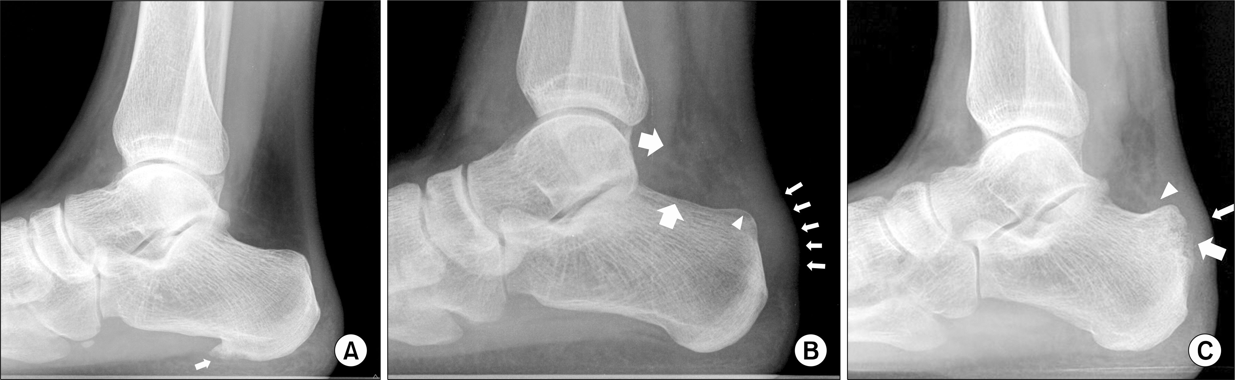
Figure 1. Lateral plain foot radiographs. (A) Subplantar erosion with an irregular spur (small arrow) in the inferior calcaneal tuberosity without any involvement soft tissue and bone of the posterior heel. (B) Focal involvement of soft tissues of the posterior heel such as blurred Kager's triangle (large arrows), swelling of the posterior soft tissue shadow (small arrows) and obliteration of the retrocalcaneal recess (arrowhead), which are distinctively comparable to posterior heel of (A). (C) Concomitant involvement of bone and soft tissue of the posterior heel such as blurred Kager's triangle, erosion of the posterior calcaneal tuberosity (large arrow), swelling of the posterior soft tissue shadow (small arrow) and obliteration of the retrocalcaneal recess (arrowhead).
Reference
-
1. Gerster JC, Vischer TL, Bennani A, Fallet GH. The painful heel. Comparative study in rheumatoid arthritis, ankylosing spondylitis, Reiter's syndrome, and generalized osteoarthrosis. Ann Rheum Dis. 1977; 36:343–8.
Article2. Dougados M, van der Linden S, Juhlin R, Huitfeldt B, Amor B, Calin A, et al. The European Spondylarthropathy Study Group preliminary criteria for the classification of spondylarthropathy. Arthritis Rheum. 1991; 34:1218–27.
Article3. Spadaro A, Iagnocco A, Perrotta FM, Modesti M, Scarno A, Valesini G. Clinical and ultrasonography assessment of peripheral enthesitis in ankylosing spondylitis. Rheumatology (Oxford). 2011; 50:2080–6.
Article4. Mason RM, Murray RS, Oates JK, Young AC. A comparative radiological study of Reiter's disease, rheumatoid arthritis and ankylosing spondylitis. J Bone Joint Surg Br. 1959; 41-B:137–48.
Article5. Gibbon WW, Cassar-Pullicino VN. Heel pain. Ann Rheum Dis. 1994; 53:344–8.
Article6. Rudwaleit M, Khan MA, Sieper J. The challenge of diagnosis and classification in early ankylosing spondylitis: do we need new criteria? Arthritis Rheum. 2005; 52:1000–8.
Article7. Ory PA. Radiography in the assessment of musculoskeletal conditions. Best Pract Res Clin Rheumatol. 2003; 17:495–512.
Article8. Resnick D, Feingold ML, Curd J, Niwayama G, Goergen TG. Calcaneal abnormalities in articular disorders. Rheumatoid arthritis, ankylosing spondylitis, psoriatic arthritis, and Reiter syndrome. Radiology. 1977; 125:355–66.9. López-Bote JP, Humbria-Mendiola A, Ossorio-Castellanos C, Padrón-Pérez M, Sabando-Suárez P. The calcaneus in ankylosing spondylitis. A radiographic study of 43 patients. Scand J Rheumatol. 1989; 18:143–8.
Article10. van der Linden S, Valkenburg HA, Cats A. Evaluation of diagnostic criteria for ankylosing spondylitis. A proposal for modification of the New York criteria. Arthritis Rheum. 1984; 27:361–8.11. van Sterkenburg MN, Muller B, Maas M, Sierevelt IN, van Dijk CN. Appearance of the weight-bearing lateral radiograph in retrocalcaneal bursitis. Acta Orthop. 2010; 81:387–90.
Article12. Landis JR, Koch GG. The measurement of observer agreement for categorical data. Biometrics. 1977; 33:159–74.
Article13. Riley MJ, Ansell BM, Bywaters EG. Radiological manifestations of ankylosing spondylitis according to age at onset. Ann Rheum Dis. 1971; 30:138–48.
Article14. Genc H, Cakit BD, Tuncbilek I, Erdem HR. Ultrasonographic evaluation of tendons and enthesal sites in rheumatoid arthritis: comparison with ankylosing spondylitis and healthy subjects. Clin Rheumatol. 2005; 24:272–7.
Article15. Fernández del Vallado P, Gálvez Failde JM, Gijón Baños J, Beltrán Gutiérrez J. Erosive calcaneitis as an early diagnostic sign in ankylosing spondylitis. Rev Clin Esp. 1967; 105:367–72.16. Bansal GJ. Digital radiography. A comparison with modern conventional imaging. Postgrad Med J. 2006; 82:425–8.
Article17. D'Agostino MA, Olivieri I. Enthesitis. Best Pract Res Clin Rheumatol. 2006; 20:473–86.18. Murphey MD. Computed radiography in musculoskeletal imaging. Semin Roentgenol. 1997; 32:64–76.
Article19. Freedman M, Steller D. Digital radiography of the musculoskeletal system: the optimal image. J Digit Imaging. 1995; 8(1 Suppl 1):37–42.
Article20. Goodman LR, Shanser JD. The pre-Achilles fat pad: An aid to early diagnosis of local or systemic disease. Skeletal Radiology. 1977; 2:81–6.
Article
- Full Text Links
-
- Actions
-
Cited
- CITED
-
- Close
- Share
-
- Similar articles
-
- Sacroiliitis in Ankylosing Spondylitis: Comparison with Multidetector Row CT and Plain Radiography
- MRI of Cauda Equina Syndrome in Ankylosing Spondylitis: A Case Report
- Ankylosing Spondylitis: Prevention And Surgical Correction Of Deformity
- Clinieal Values of Single Photon Emission Computed Tomography ( SPECT ) in Ankylosing Spondylitis
- Clinical features of ankylosing spondylitis
