Changes in Strain Pattern and Exercise Capacity after Transcatheter Closure of Atrial Septal Defects
- Affiliations
-
- 1Division of Pediatric Cardiology, Department of Pediatrics, Congenital Heart Disease Center, Severance Cardiovascular Hospital, Yonsei University College of Medicine, Seoul, Korea. pednk@yuhs.ac
- 2Department of Pediatrics, National Health Insurance Service Ilsan Hospital, Goyang, Korea.
- KMID: 2377466
- DOI: http://doi.org/10.4070/kcj.2016.0205
Abstract
- BACKGROUND AND OBJECTIVES
Assessment of left ventricle (LV) function by using strain and strain rate is popular in the clinical setting. However, the use of these echocardiographic tools in assessing right ventricle (RV) failure, and the manner in which they both reflect the functional capacity of the patient, remains poorly understood. This study aimed to investigate the change in exercise capacity and strain between before and (1 month) after the transcatheter closure of atrial septal defects (ASDs).
SUBJECTS AND METHODS
Thirty patients who underwent transcatheter closure of ASD between May 2014 and June 2015 at the Division of Pediatric Cardiology, Severance Cardiovascular Hospital, were enrolled. We compared and analyzed the results of the following examinations, before and (1 month) after the procedure: echocardiography, cardiopulmonary exercise test (CPET), and N-terminal pro-brain natriuretic peptide level.
RESULTS
There were no mortalities, and the male-to-female ratio was 1:2. The mean defect size was 22.3±4.9 mm; the mean Qp/Qs ratio, 2.1±0.5; and the mean device size, 22.3±4.9 mm. Changes in global RV longitudinal (GRVL) strain and LV torsion were measured echocardiographically. Exercise capacity improved from 7.7±1.2 to 8.7±1.8 metabolic equivalents (p=0.001). These findings correlated to the change in GRVL strain (p=0.03).
CONCLUSION
The average exercise capacity increased after device closure of ASD. The change in strain was evident on echocardiography, especially for GRVL strain and LV torsion. Further studies comparing CPET and strain in various patients may show increased exercise capacity in patients with improved RV function.
MeSH Terms
Figure
-
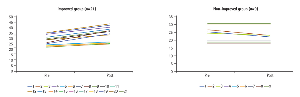
Fig. 1 Individual exercise capacity changed in each group. The improved group was defined as patients with improved exercise capacity before and after the procedure. The non-improved group was defined as patients with decreased or unchanged exercise capacity before and after the procedure.
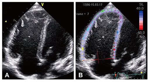
Fig. 2 Longitudinal strain of the right ventricle, in the four-chamber view. (A) Two-dimensional image of the four-chamber view. (B) Right ventricle strain on the speckle tracking image, semi-automatically divided into six segments (basal, mid, and apical segments of the septum and those of the right ventricle free wall).
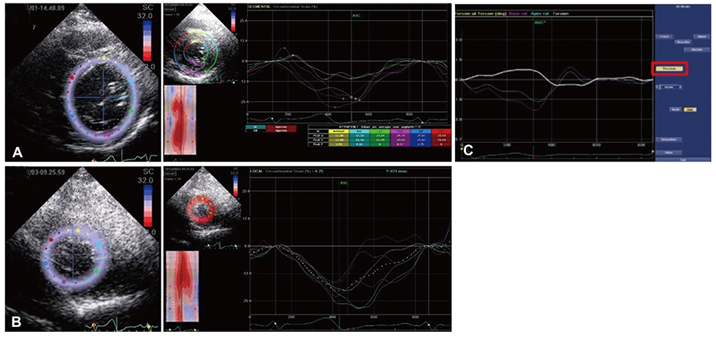
Fig. 3 LV torsion. (A) Parasternal short axis view, mitral level. (B) Parasternal short axis view, apical level. (C) LV torsion was obtained from (A) and (B), and calculated by using the EchoPAC system (GE Medical Systems, Milwaukee, WI, USA). MPV: mean platelet volume, BNP: brain natriuretic peptide, LV: left ventricle.
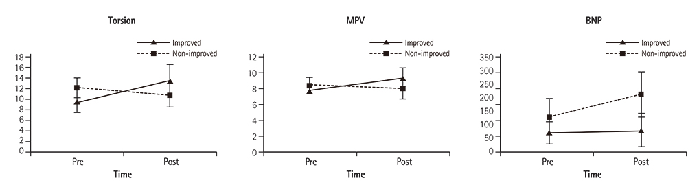
Fig. 4 Mean profile graphs of the differences between groups. MPV: mean platelet volume, BNP: brain natriuretic peptide.
Cited by 2 articles
-
Procedural, Early and Long-Term Outcomes after Transcatheter Atrial Septal Defects Closure: Comparison between Large and Very Large Atrial Septal Defect Groups
So-Ick Jang
Korean Circ J. 2019;49(10):987-989. doi: 10.4070/kcj.2019.0165.Comparison of Predicted Exercise Capacity Equations in Adult Korean Subjects
Daehyun Jeong, Yeon-Mok Oh, Sei Won Lee, Sang-Do Lee, Jae Seung Lee
J Korean Med Sci. 2022;37(14):e113. doi: 10.3346/jkms.2022.37.e113.
Reference
-
1. D'Hooge J, Herbots L, Sutherland GR. Quantitative assessment of intrinsic regional myocardial deformation by Doppler strain rate echocardiography in humans. Circulation. 2003; 107:e49. author reply e49.2. Mooman-Smook JC, Brink PA. Striving towards the ideal cardiac functional assessment strategy: the contribution of tissue Doppler, strain and strain rate imaging. Cardiovasc J Afr. 2007; 18:387–392.3. Baur LH. Strain and strain rate imaging: a promising tool for evaluation of ventricular function. Int J Cardiovasc Imaging. 2008; 24:493–494.4. Pislaru C, Pellikka PA. Tissue Doppler and strain-rate imaging in cardiac ultrasound imaging: valuable tools or expensive ornaments? Expert Rev Cardiovasc Ther. 2005; 3:1–4.5. Vitarelli A, Conde Y, Cimino E, et al. Assessment of right ventricular function by strain rate imaging in chronic obstructive pulmonary disease. Eur Respir J. 2006; 27:268–275.6. Chen J, Cao T, Duan Y, Yuan L, Wang Z. Velocity vector imaging in assessing myocardial systolic function of hypertensive patients with left ventricular hypertrophy. Can J Cardiol. 2007; 23:957–961.7. Takeuchi M, Borden WB, Nakai H, et al. Reduced and delayed untwisting of the left ventricle in patients with hypertension and left ventricular hypertrophy: a study using two-dimensional speckle tracking imaging. Eur Heart J. 2007; 28:2756–2762.8. Gorcsan J 3rd, Suffoletto MS. The role of tissue Doppler and strain imaging in predicting response to CRT. Europace. 2008; 10:Suppl 3. iii80–iii87.9. Valsangiacomo Buechel ER, Mertens LL. Imaging the right heart: the use of integrated multimodality imaging. Eur Heart J. 2012; 33:949–960.10. Mertens LL, Friedberg MK. Imaging the right ventricle--current state of the art. Nat Rev Cardiol. 2010; 7:551–563.11. Brochu MC, Baril JF, Dore A, Juneau M, De Guise P, Mercier LA. Improvement in exercise capacity in asymptomatic and mildly symptomatic adults after atrial septal defect percutaneous closure. Circulation. 2002; 106:1821–1826.12. Su CT, Sung TY, Lin KL, Wang JL, Yang AL. Lower exercise capacity in children with asymptomatic atrial septal defect associated with circulatory impairment. Chin J Physiol. 2013; 56:110–116.13. Weber M, Neumann T, Rau M, et al. Cardiopulmonary exercise capacity increases after interventional ASD-closure. Z Kardiol. 2004; 93:209–215.14. Kaya MG, Elcik D, Akpek M, et al. Mean platelet volume levels predict pulmonary artery hypertension in patients with atrial septal defect. Acta Cardiol. 2014; 69:161–166.15. Elsheikh RG, Hegab M, Szatmari A. NT-proBNP correlated with strain and strain rate imaging of the right ventricle before and after transcatheter closure of atrial septal defects. J Saudi Heart Assoc. 2013; 25:3–8.16. Kaminsky LA, Whaley MH. Evaluation of a new standardized ramp protocol: the BSU/Bruce Ramp protocol. J Cardiopulm Rehabil. 1998; 18:438–444.17. Friedberg MK, Mertens L. Tissue velocities, strain, and strain rate for echocardiographic assessment of ventricular function in congenital heart disease. Eur J Echocardiogr. 2009; 10:585–593.18. Bussadori C, Moreo A, Di Donato M, et al. A new 2D-based method for myocardial velocity strain and strain rate quantification in a normal adult and paediatric population: assessment of reference values. Cardiovasc Ultrasound. 2009; 7:8.19. Nagaya N, Nishikimi T, Okano Y, et al. Plasma brain natriuretic peptide levels increase in proportion to the extent of right ventricular dysfunction in pulmonary hypertension. J Am Coll Cardiol. 1998; 31:202–208.20. Nagaya N, Nishikimi T, Uematsu M, et al. Secretion patterns of brain natriuretic peptide and atrial natriuretic peptide in patients with or without pulmonary hypertension complicating atrial septal defect. Am Heart J. 1998; 136:297–301.21. Dong L, Zhang F, Shu X, et al. Left ventricular torsional deformation in patients undergoing transcatheter closure of secundum atrial septal defect. Int J Cardiovasc Imaging. 2009; 25:479–486.22. Wu ET, Akagi T, Taniguchi M, et al. Differences in right and left ventricular remodeling after transcatheter closure of atrial septal defect among adults. Catheter Cardiovasc Interv. 2007; 69:866–871.23. Giardini A, Donti A, Formigari R, et al. Determinants of cardiopulmonary functional improvement after transcatheter atrial septal defect closure in asymptomatic adults. J Am Coll Cardiol. 2004; 43:1886–1891.24. Li X, Chen C, Gan F, Wang Y, Ding L, Hua W. Plasma NT pro-BNP, hs-CRP and big-ET levels at admission as prognostic markers of survival in hospitalized patients with dilated cardiomyopathy: a single-center cohort study. BMC Cardiovasc Disord. 2014; 14:67.
- Full Text Links
-
- Actions
-
Cited
- CITED
-
- Close
- Share
-
- Similar articles
-
- Comprehensive understanding of atrial septal defects by imaging studies for successful transcatheter closure
- Procedural, Early and Long-Term Outcomes after Transcatheter Atrial Septal Defects Closure: Comparison between Large and Very Large Atrial Septal Defect Groups
- Emergent Surgical Intervention for Embolization of Atrial Septal Defect Closure Device
- Transcatheter Closure of Secundum Atrial Septal Defect with the Amplatzer Septal Occluder
- Outcome of Transcatheter Closure of Oval Shaped Atrial Septal Defect with Amplatzer Septal Occluder
