A large osteoid osteoma of the mandibular condyle causing conductive hearing loss: a case report and review of literature
- Affiliations
-
- 1Richardsons Dental and Craniofacial Hospital, Nagercoil, India. sunilrichardson145@gmail.com
- KMID: 2377014
- DOI: http://doi.org/10.5125/jkaoms.2017.43.2.106
Abstract
- Osteoid osteomas are benign skeletal neoplasms that are commonly encountered in the bones of the lower extremities, but are exceedingly rare in jaw bones with a prevalence of less than 1%. This unique clinical entity is usually seen in younger individuals, with nocturnal pain and swelling as its characteristic clinical manifestations. The size of the lesion is rarely found to be more than 2 cm. We hereby report a rare case of osteoid osteoma originating from the neck of the mandibular condyle that grew to large enough proportions to result in conductive hearing loss in addition to pain, swelling and restricted mouth opening. In addition, an effort has been made to review all the documented cases of osteoid osteomas of the jaws that have been published in the literature thus far.
Keyword
MeSH Terms
Figure
-
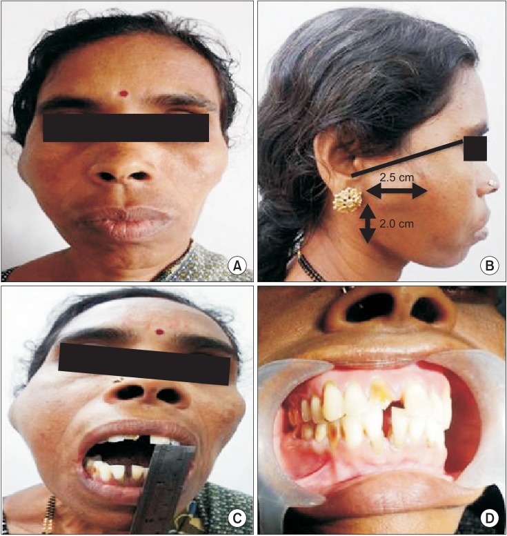
Fig. 1 Preoperative photographs of the patient. A. Frontal view. B. Lateral view with clinical extensions of the lesion. C. Photograph depicting restricted mouth opening. D. Intraoral view.
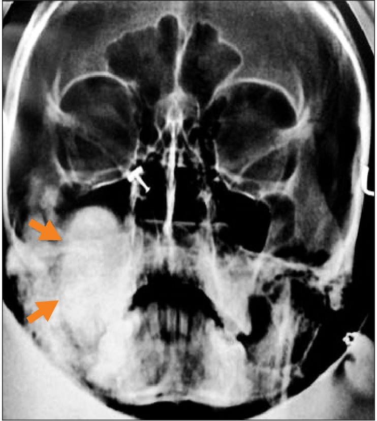
Fig. 2 Preoperative plain radiograph (Waters view) showing a well defined radio-opacity (arrows).
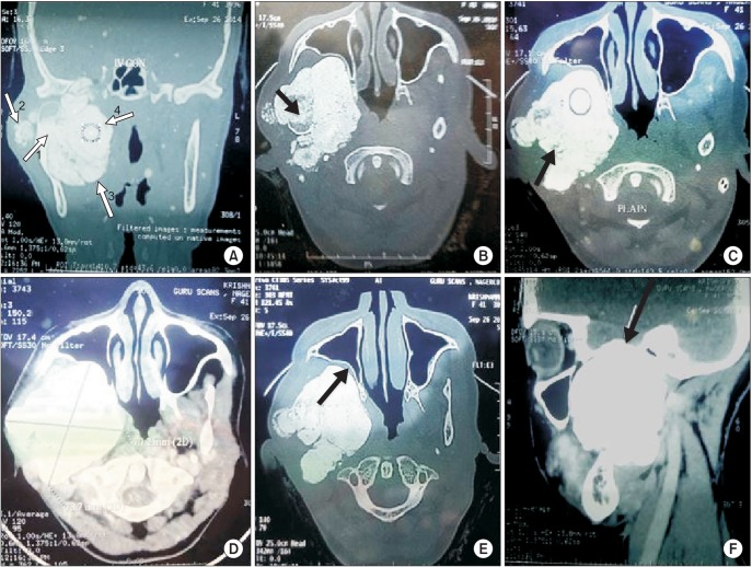
Fig. 3 Computed tomographic (CT) views of the case (plain and contrast enhanced). A. CT view showing the origin of the lesion from the neck of the condyle (arrow 1), nidus (arrow 2), and extent of the lesion (arrow 3 and 4). B. Origin of the lesion from the condylar neck (black arrow). C. Origin of the lesion from the condylar neck (black arrow). D. Contrast enhanced view showing the dimensions of the lesion (2D). E. View showing indentation of the posterior maxillary sinus wall (black arrow). F. View showing lesion extension intracranially into the right middle cranial fossa (black arrow).
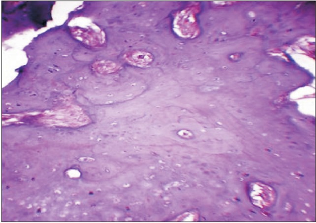
Fig. 4 Histopathologic picture showing features consistent with osteoid osteoma.
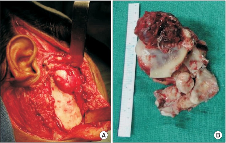
Fig. 5 Intraoperative photographs. A. Incision planning and exposure of the lesion. B. Excised specimen in toto.
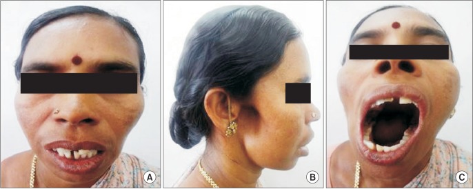
Fig. 6 Postoperative clinical photographs at 1 year. A. Frontal view. B. Profile view showing the hollowness in the right temporomandibular joint region and a cosmetically acceptable scar. C. Photograph depicting improvement in mouth opening.
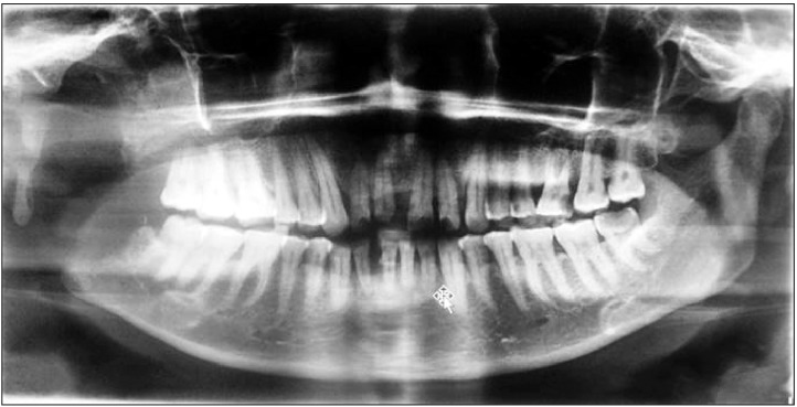
Fig. 7 Postoperative panoramic radiograph at 1 year.
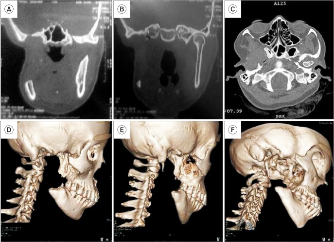
Fig. 8 Postoperative plain computed tomographic (CT) scan at 1 year with three-dimensional (3D) reconstructive images showing no recurrence. A-C. CT views depicting no recurrence. D-F. 3D reconstructive images.
Reference
-
1. Jaffe HL. Osteoid osteoma: a benign osteoblastic tumor composed of osteoid and atypical bone. Arch Surg. 1935; 31:709–728.2. Ida M, Kurabayashi T, Takahashi Y, Takagi M, Sasaki T. Osteoid osteoma in the mandible. Dentomaxillofac Radiol. 2002; 31:385–387. PMID: 12424638.
Article3. Yang C, Qiu WL. Osteoid osteoma of the eminence of the temporomandibular joint. Br J Oral Maxillofac Surg. 2001; 39:404–406. PMID: 11601826.
Article4. Singh A, Solomon MC. Osteoid osteoma of the mandible: a case report with review of the literature. J Dent Sci. 2012; DOI: 10.1016/j.jds.2012.10.002. [Epub ahead of print].
Article5. An SY, Shin HI, Choi KS, Park JW, Kim YG, Benavides E, et al. Unusual osteoid osteoma of the mandible: report of case and review of the literature. Oral Surg Oral Med Oral Pathol Oral Radiol. 2013; 116:e134–e140. PMID: 23849381.
Article6. Foss EL, Dockerty MB, Good CA. Osteoid osteoma of the mandible; report of a case. Cancer. 1955; 8:592–594. PMID: 14379148.7. Gupta OP, Jain RK, Agarwal MK, Khanna S, Shrivastava A. Osteoid osteoma of the mandible. Ear Nose Throat J. 1985; 64:206. 208. PMID: 3996270.8. Tochihara S, Sato T, Yamamoto H, Asada K, Ishibashi K. Osteoid osteoma in mandibular condyle. Int J Oral Maxillofac Surg. 2001; 30:455–457. PMID: 11720052.
Article9. Chaudhary M, Kulkarni M. Osteoid osteoma of the mandible. J Oral Maxillofac Pathol. 2007; 11:52–55.10. Rahsepar B, Nikgoo A, Fatemitabar SA. Osteoid osteoma of subcondylar region: case report and review of the literature. J Oral Maxillofac Surg. 2009; 67:888–893. PMID: 19304051.
Article11. Karandikar S, Thakur G, Tijare M, Shreenivas K, Agrawal K. Osteoid osteoma of mandible. BMJ Case Rep. 2011; 2011:bcr1020114886.
Article12. Mohammed I, Jannan NA, Elrmali A. Osteoid osteoma associated with the teeth: unusual presentation. Int J Oral Maxillofac Surg. 2013; 42:298–302. PMID: 22658497.
Article13. Barei DP, Moreau G, Scarborough MT, Neel MD. Percutaneous radiofrequency ablation of osteoid osteoma. Clin Orthop Relat Res. 2000; (373):115–124.
Article14. Pritchard JE, Mckay JW. Osteoid osteoma. Can Med Assoc J. 1948; 58:567–575. PMID: 18862254.15. Alvares Capelozza AL, Gião Dezotti MS, Casati Alvares L, Negrão Fleury R, Sant'Ana E. Osteoblastoma of the mandible: systematic review of the literature and report of a case. Dentomaxillofac Radiol. 2005; 34:1–8. PMID: 15709098.
Article16. Rawal YB, Angiero F, Allen CM, Kalmar JR, Sedghizadeh PP, Steinhilber AM. Gnathic osteoblastoma: clinicopathologic review of seven cases with long-term follow-up. Oral Oncol. 2006; 42:123–130. PMID: 16129654.
Article17. Jones AC, Prihoda TJ, Kacher JE, Odingo NA, Freedman PD. Osteoblastoma of the maxilla and mandible: a report of 24 cases, review of the literature, and discussion of its relationship to osteoid osteoma of the jaws. Oral Surg Oral Med Oral Pathol Oral Radiol Endod. 2006; 102:639–650. PMID: 17052641.
Article18. Goldberg VM, Jacobs B. Osteoid osteoma of the hip in children. Clin Orthop Relat Res. 1975; (106):41–47.
Article19. Sim FH, Dahlin CD, Beabout JW. Osteoid-osteoma: diagnostic problems. J Bone Joint Surg Am. 1975; 57:154–159. PMID: 1112841.
