A case of lymphocytic variant hypereosinophilic syndrome with sub-diagnostic systemic mastocytosis
- Affiliations
-
- 1Department of Leukemia, The University of Texas, M.D. Anderson Cancer Center, Houston, Texas, USA. zestrov@mdanderson.org
- 2Department of Hematopathology, The University of Texas, M.D. Anderson Cancer Center, Houston, Texas, USA.
- KMID: 2375210
- DOI: http://doi.org/10.5045/br.2017.52.1.71
Abstract
- No abstract available.
Figure
-
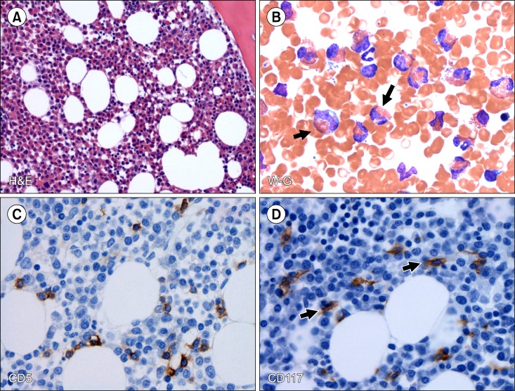
Fig. 1 Morphologic and immunophenotypic features of the bone marrow biopsy and aspirate. (A) Bone marrow core biopsy shows hypercellular bone marrow with trilineage hematopoiesis and markedly increased eosinophils (H&E, ×200). (B) Bone marrow aspirate smear shows increased eosinophils, black arrows (Wright-Giemsa, ×500). (C) Immunohistochemical stain for CD5 shows a few scattered T cells (CD5, ×500). (D) Immunohistochemical stain for CD117 shows slightly increased atypical spindle-shaped mast cells (black arrows) with interstitial distribution (500) representing subdiagnostic mastocytosis.
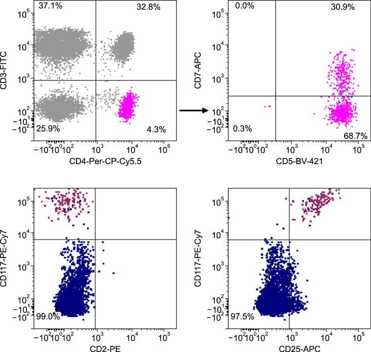
Fig. 2 Flow cytometry analysis of bone marrow aspirate detected a minor population of aberrant T cells (about 0.52% of total cells) coexpressing CD4, CD5 with partial loss of CD7 (upper panel) and CD2 expression (not shown), and an aberrant population of cells coexpressing CD25 and CD117 mast cells (0.04%) (lower panel).
Reference
-
1. Pardanani A, Chen D, Abdelrahman RA, et al. Clonal mast cell disease not meeting WHO criteria for diagnosis of mastocytosis: clinicopathologic features and comparison with indolent mastocytosis. Leukemia. 2013; 27:2091–2094. PMID: 23896642.
Article2. Helbig G, Wieczorkiewicz A, Dziaczkowska-Suszek J, Majewski M, Kyrcz-Krzemien S. T-cell abnormalities are present at high frequencies in patients with hypereosinophilic syndrome. Haematologica. 2009; 94:1236–1241. PMID: 19734416.
Article3. Christoforidou A, Kotsianidis I, Margaritis D, Anastasiadis A, Douvali E, Tsatalas C. Long-term remission of lymphocytic hypereosinophilic syndrome with imatinib mesylate. Am J Hematol. 2012; 87:131–132.
Article4. Roufosse F. Peripheral T-cell lymphoma developing after diagnosis of lymphocytic variant hypereosinophilic syndrome: misdiagnosed lymphoma or natural disease progression? Oral Surg Oral Med Oral Pathol Oral Radiol. 2014; 118:506–510.
Article5. Wiednig M, Beham-Schmid C, Kranzelbinder B, Aberer E. Clonal mast cell proliferation in pruriginous skin in hypereosinophilic syndrome. Dermatology. 2013; 227:67–71. PMID: 24008407.
Article
- Full Text Links
-
- Actions
-
Cited
- CITED
-
- Close
- Share
-
- Similar articles
-
- A Case of Adult-onset Urticaria Pigmentosa with Bone Involvement
- A Case of the Idiopathic Hypereosinophilic Syndrome Evolving to Malignant Lymphoma
- Solitary mastocytoma presenting at birth
- Idiopathic Hypereosinophilic Syndrome Involving Thoracic Spine
- A Case of Huge Left Ventricular Thrombus Associated with Hypereosinophilic Syndrome
