Esophageal Hemangioma Treated by Endoscopic Mucosal Resection: A Case Report and Review of the Literature
- Affiliations
-
- 1Division of Gastroenterology, Department of Internal Medicine, Korea University College of Medicine, Seoul, Korea. sungwoojung@korea.ac.kr
- KMID: 2373357
- DOI: http://doi.org/10.4166/kjg.2015.66.5.277
Abstract
- Hemangioma of the esophagus is a rare form of benign esophageal tumor. It usually presents as a single lesion located in the lower third of the esophagus and is mostly asymptomatic. However, it may occasionally cause hematemesis and/or obstruction. Surgical resection is the conventional treatment modality for managing esophageal hemangioma, but less invasive approaches such as endoscopic therapy are recently becoming more widely employed. Herein, we report a case of a 54-year-old man who presented with an esophageal hemangioma that was successfully treated by endoscopic mucosal resection without any complications.
Keyword
MeSH Terms
Figure
-
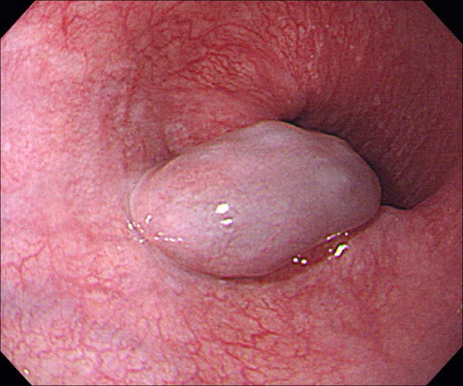
Fig. 1. Endoscopic image showing a bluish soft round protruding submucosal mass in the lower esophagus.
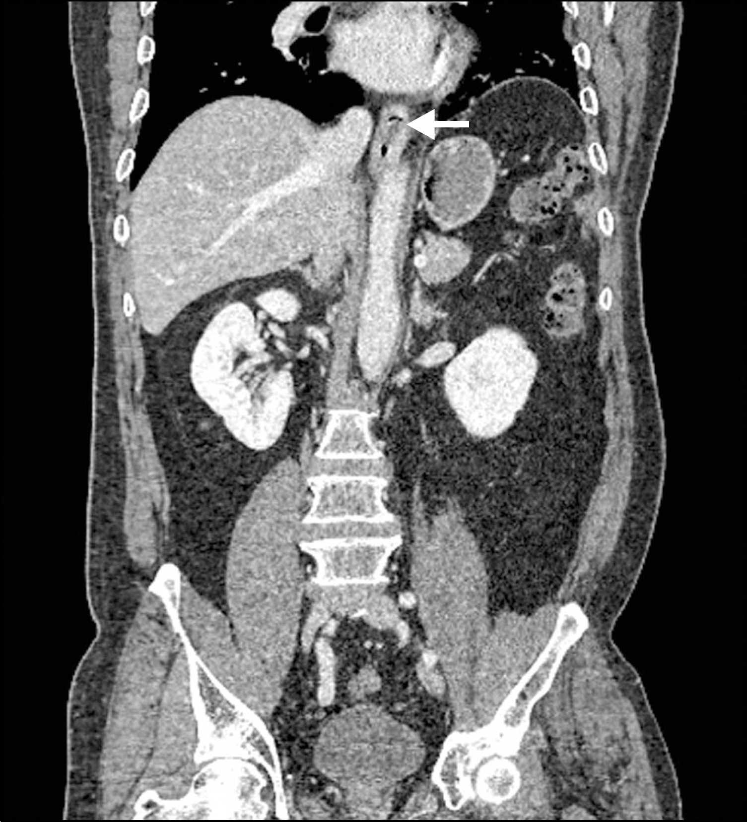
Fig. 2. CT finding. A poorly enhancing mass (arrow) is observed within the lower esophageal lumen.
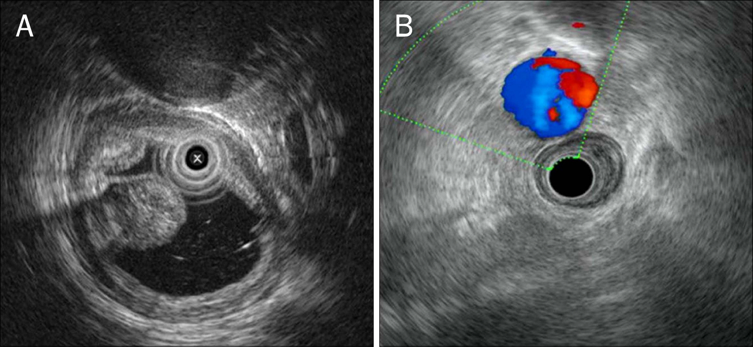
Fig. 3. EUS findings. (A) The tumor is located in the submucosal layer and shows heterogenous echogenicity that is well separated from adjacent tissue. (B) It has no continuity with adjacent vessels.
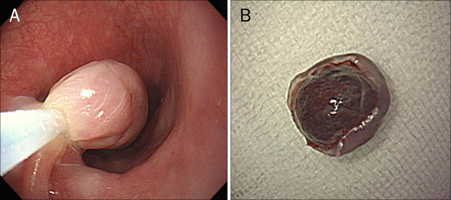
Fig. 4. (A) The lesion is being resected by conventional endoscopic mucosal resection technique after injecting saline into the base of the mass. (B) The resected specimen is a dark red colored mass that measures 14×14 mm in size.
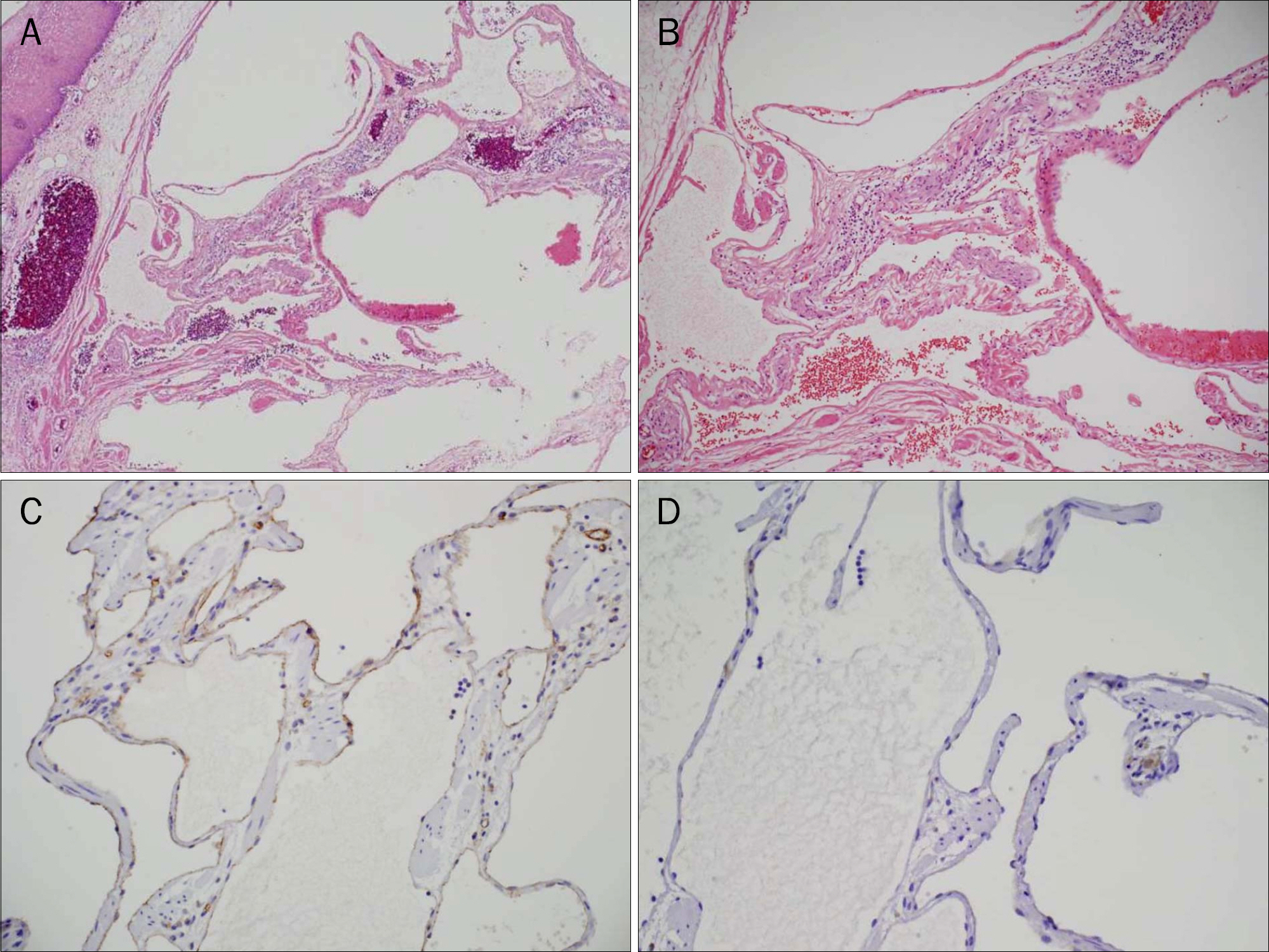
Fig. 5. Histopathologic findings. (A) Dilated blood vessels are surrounded by flat endothelial cells in the submucosa (H&E, ×40). (B) Dilated vessels lined by flattened endothelium show communication with each other (H&E, ×100). (C) The endothelial cells are positive for CD-31 and (D) negative for D2–40 on immunohistochemistry stain (×200).
Reference
-
References
1. Choong CK, Meyers BF. Benign esophageal tumors: introduction, incidence, classification, and clinical features. Semin Thorac Cardiovasc Surg. 2003; 15:3–8.
Article2. Rice TW. Benign esophageal tumors: esophagoscopy and endoscopic esophageal ultrasound. Semin Thorac Cardiovasc Surg. 2003; 15:20–26.
Article3. Sogabe M, Taniki T, Fukui Y, et al. A patient with esophageal hemangioma treated by endoscopic mucosal resection: a case report and review of the literature. J Med Invest. 2006; 53:177–182.
Article4. Park SW, Moon JS, Lee SR, et al. A case of esophageal hemangioma treated by endoscopic mucosal resection. Korean J Gastrointest Endosc. 2010; 40:244–248.5. Gentry RW, Dockerty MB, Glagett OT. Vascular malformations and vascular tumors of the gastrointestinal tract. Surg Gynecol Obstet. 1949; 88:281–323.6. Plachta A. Benign tumors of the esophagus. Review of literature and report of 99 cases. Am J Gastroenterol. 1962; 38:639–652.7. Shigemitsu K, Naomoto Y, Yamatsuji T, et al. Esophageal hemangioma successfully treated by fulguration using potassium titanyl phosphate/yttrium aluminum garnet (KTP/YAG) laser: a case report. Dis Esophagus. 2000; 13:161–164.
Article8. Rajoriya N, D'costa H, Gupta P, Ellis AJ. An unusual cause of dyspepsia: oesophageal cavernous haemangioma. QJM. 2010; 103:791–793.
Article9. Hanel K, Talley NA, Hunt DR. Hemangioma of the esophagus: an unusual cause of upper gastrointestinal bleeding. Dig Dis Sci. 1981; 26:257–263.
Article10. Tsai SJ, Lin CC, Chang CW, et al. Benign esophageal lesions: endoscopic and pathologic features. World J Gastroenterol. 2015; 21:1091–1098.
Article11. Araki K, Ohno S, Egashira A, et al. Esophageal hemangioma: a case report and review of the literature. Hepatogastroenterology. 1999; 46:3148–3154.12. Ghiatas AA, Chopra S, Escobar B, Esola CC, Chintapalli K, Dodd GD 3rd. Esophageal hemangioma. Eur Radiol. 1997; 7:1062–1063.
Article13. Lee HJ, Kim YT, Sung SW, Kim JH. A case of long segment my-omectomy for the treatment of esophageal hemangioma. Korean J Thorac Cardiovasc Surg. 2003; 36:206–210.14. Raza W, Nasir H, Ilahi F, Zafar Z. Esophageal haemangioma: a case report and review of literature. J Pak Med Assoc. 2010; 60:310–311.15. Aoki T, Okagawa K, Uemura Y, et al. Successful treatment of an esophageal hemangioma by endoscopic injection sclerotherapy: report of a case. Surg Today. 1997; 27:450–452.
Article16. Shin S, Choi YS, Shim YM, Kim HK, Kim K, Kim J. Enucleation of esophageal submucosal tumors: a single institution's experience. Ann Thorac Surg. 2014; 97:454–459.
Article
- Full Text Links
-
- Actions
-
Cited
- CITED
-
- Close
- Share
-
- Similar articles
-
- A Case of Esophageal Hemangioma Treated by Endoscopic Mucosal Resection
- Successful Endoscopic Mucosal Resection of a Low Esophageal Carcinoid Tumor
- A Case of Early Esophageal Cancer Treated by Endoscopic Mucosal Resection Using a EEMR Tube
- A Case of High Grade Dysplasia Arising from Barrett's Esophagus Treated with Endoscopic Mucosal Resection
- A Case of Long Segment Myomectomy for the Treatment of Esophageal Hemangioma
