Lymphoepithelial Cyst of the Pancreas
- Affiliations
-
- 1Division of Gastroenterology and Hepatology, Department of Internal Medicine, Korea University Ansan Hospital, Korea University College of Medicine, Ansan, Korea. sean4h@korea.ac.kr
- KMID: 2373242
- DOI: http://doi.org/10.4166/kjg.2015.65.6.379
Abstract
- No abstract available.
MeSH Terms
Figure
-
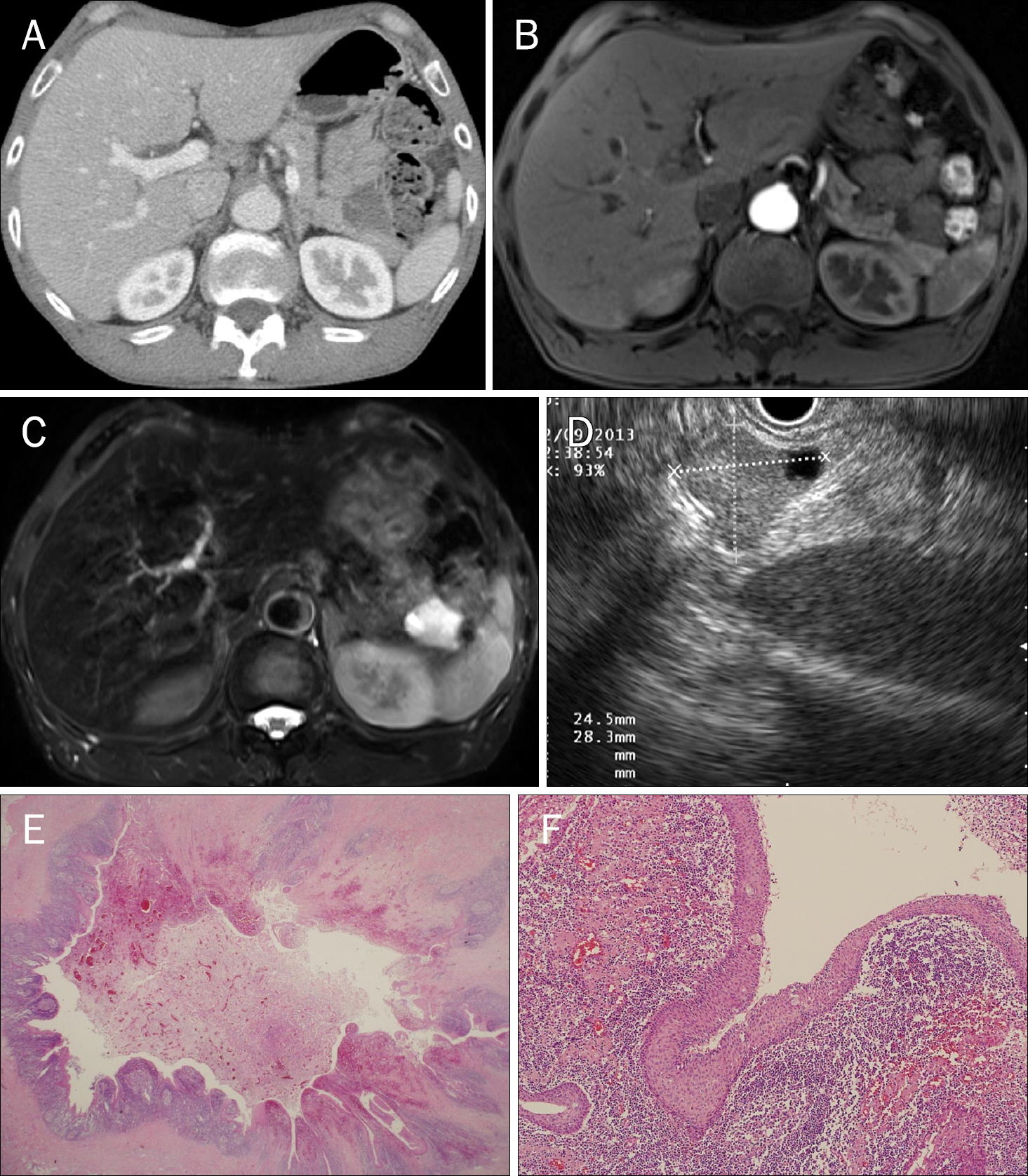
Fig. 1. (A) Abdominal CT scan shows a non-enhancing septated cystic mass at pancreas tail. (B) T1-weighted image on MRCP shows a septated cystic mass with slightly high signal intensity of the larger chamber. (C) T2-weighted image of MRCP shows a septated cystic mass with slightly low signal intensity of the larger chamber. (D) Endoscopic ultrasound shows a heterogeneously isoechoic cystic mass with daughter cystic lesion. (E) A submucosal cyst lined with stratified squamous epithelium is seen and in some areas, the lining epithelium is attenuated or denuded with erosion (H&E stain, ×15). (F) There is an aggregation of lymphocytes forming a subepithelial rim of lymphoid tissue (H&E stain, ×200).
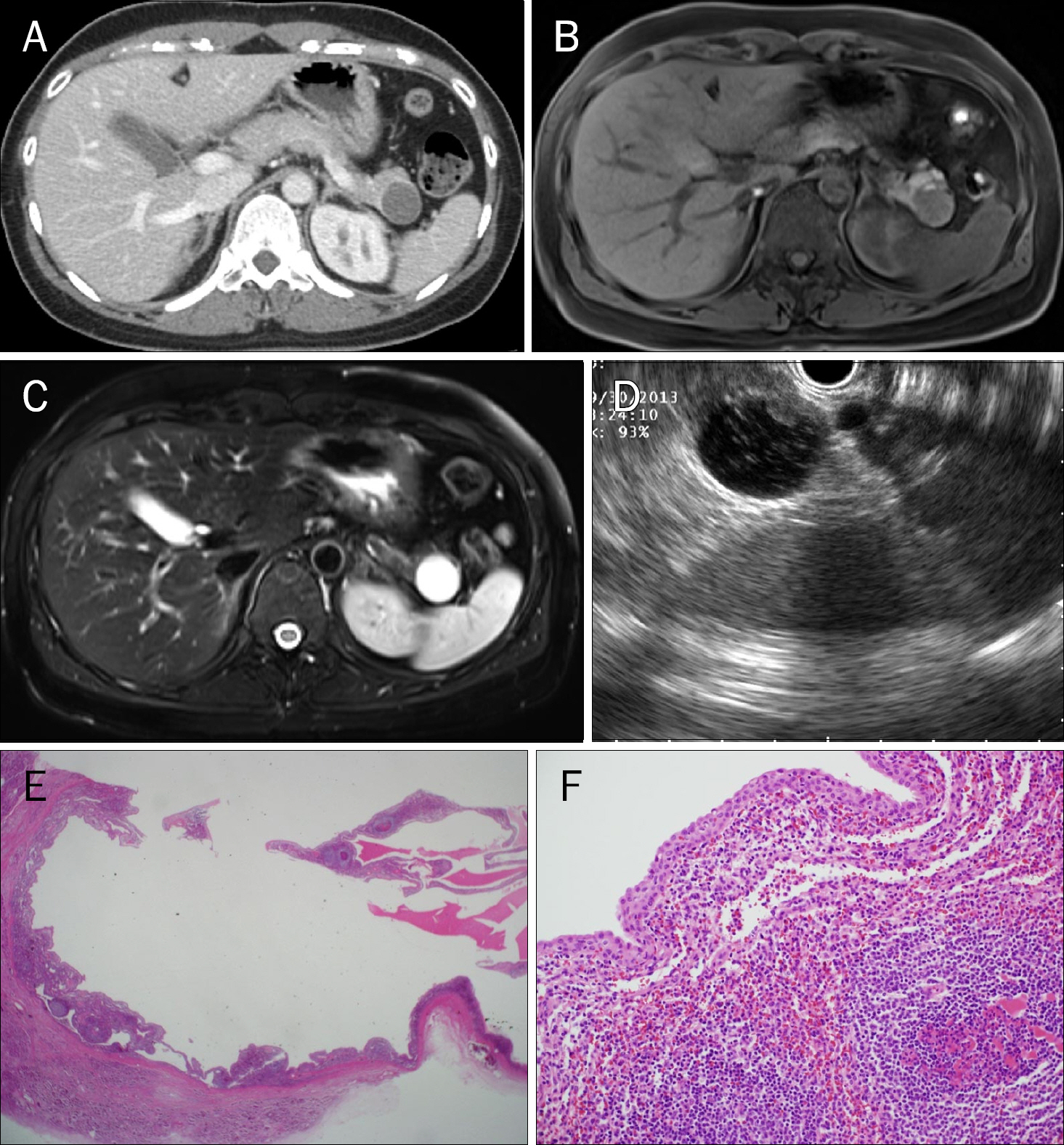
Fig. 2. (A) Abdominal CT shows a peripherally enhancing cystic mass at pancreas tail. (B) T1-weighted image of MRCP shows a well-defined cystic mass with focal area of high signal intensity in anterior wall of the mass. (C) T2-weighted image of MRCP shows a cystic mass with high signal intensity with focal wall thickening. (D) Endoscopic ultrasound shows floating debris within the cystic mass along with focal wall thickening. (E) About 2.0 cm sized cyst is seen within the pancreas (H&E stain, ×15). (F) The lining epithelium of the cyst is squamous epithelium and there are abundant lymphocytes accompanied by germinal center formation in the cyst wall (H&E stain, ×200).
Reference
-
References
1. Lüchtrath H, Schriefers KH. A pancreatic cyst with features of a so-called branchiogenic cyst. Pathologe. 1985; 6:217–219.2. Truong LD, Rangdaeng S, Jordan PH Jr. Lymphoepithelial cyst of the pancreas. Am J Surg Pathol. 1987; 11:899–903.
Article3. Liu J, Shin HJ, Rubenchik I, Lang E, Lahoti S, Staerkel GA. Cytologic features of lymphoepithelial cyst of the pancreas: two preoperatively diagnosed cases based on fine-needle aspiration. Diagn Cytopathol. 1999; 21:346–350.
Article4. Adsay NV, Hasteh F, Cheng JD, et al. Lymphoepithelial cysts of the pancreas: a report of 12 cases and a review of the literature. Mod Pathol. 2002; 15:492–501.
Article5. Hastings PR, Nance FC, Becker WF. Changing patterns in the management of pancreatic pseudocysts. Ann Surg. 1975; 181:546–551.
Article6. Fujiwara H, Kohno N, Nakaya S, Ishikawa Y. Lymphoepithelial cyst of the pancreas with sebaceous differentiation. J Gastroenterol. 2000; 35:396–401.
Article7. Bolis GB, Farabi R, Liberati F, Macciò T. Lymphoepithelial cyst of the pancreas. Report of a case diagnosed by fine needle aspiration biopsy. Acta Cytol. 1998; 42:384–386.8. Capitanich P, Iovaldi ML, Medrano M, et al. Lymphoepithelial cysts of the pancreas: case report and review of the literature. J Gastrointest Surg. 2004; 8:342–345.
Article9. Park IS, Chung YH, Choi JY, et al. Lymphoepithelial cyst of the pancreas: a case report. Korean J Med. 2009; 76(Suppl 1):S54–S58.10. Lee HS, Choi SY, Jung MK, et al. A lymphoepithelial cyst of the pancreas: A case report. Korean J Med. 2010; 78:616–619.11. Kim YH, Auh YH, Kim KW, Lee MG, Kim KS, Park SY. Lymphoepithelial cysts of the pancreas: CT and sonographic findings. Abdom Imaging. 1998; 23:185–187.
Article12. Koga H, Takayasu K, Mukai K, et al. CT of lymphoepithelial cysts of the pancreas. J Comput Assist Tomogr. 1995; 19:221–224.
Article13. Fukukura Y, Inoue H, Miyazono N, et al. Lymphoepithelial cysts of the pancreas: demonstration of lipid component using CT and MRI. J Comput Assist Tomogr. 1998; 22:311–313.14. Zou XP, Li YM, Li ZS, Xu GM. Lymphoepithelial cyst of the pancreas: a case report. Hepatobiliary Pancreat Dis Int. 2004; 3:155–157.
