Gastric Lipomatosis
- Affiliations
-
- 1Department of Surgery, Jeju National University School of Medicine, Jeju, Korea. 41056@naver.com
- 2Department of Pathology, Jeju National University School of Medicine, Jeju, Korea.
Abstract
- Gastric lipomatosis is an extremely rare condition. We present a case of a 69-year-old woman admitted with epigastric soreness. Computerized tomography (CT) revealed extrinsically compressing, fat-containing mass lesions on the entire gastric wall of the antrum and body except for the lesser curvature. A subtotal gastrectomy was performed. Pathology findings confirmed a gastric lipomatosis with multiple gastric ulcerations and extensive disruptions of the muscular layers. This case and reports of other gastric lipomatosis cases indicate that CT should be used to characterize large submucosal masses because CT can show the specific nature and extent of the disease. We believe that surgical treatment is the most appropriate treatment for symptomatic gastric lipomatosis that shows extensive gastric involvement, or when there are multiple gastric lipomas.
Keyword
Figure
-
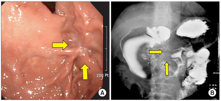
Fig. 1 Gastroscopy findings (A) and UGI study (B). Note the extrinsic compressing mass lesion on the anterior wall-greater curvature-posterior wall of the antrum and body, and the ulceration (arrows).
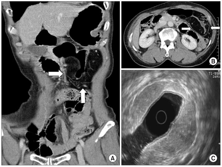
Fig. 2 CT (A and B) and EUS (C) radiology findings. Note the huge fat-containing mass lesion around the gastric wall except for the lesser curvature, from the gastric midbody to the antrum. The lesions (arrows) appeared as well-circumscribed areas of fatty density with an attenuation ranging from -70 to -120 H. The lesion originated at the submucosal layer, had a depth of about 2.7 cm and was homogeneously hyperechoic on EUS findings.
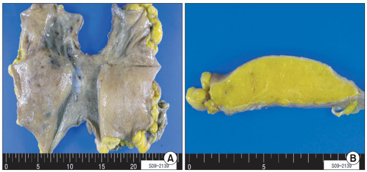
Fig. 3 Gross pathology findings of the resected specimen. (A) Examination of the intact specimen. Note the multiple nodules of deep adipose tissue without encapsulation, and the mucosal surface with several ulcers which was diffusely elevated by the submucosal mass (approximately 16×16 cm) that almost encircled the gastric wall. (B) Examination of the specimen following sectioning. Note the deep adipose tissue mainly in the submucosa, and adipose tissue diffusely disrupting the muscular wall.
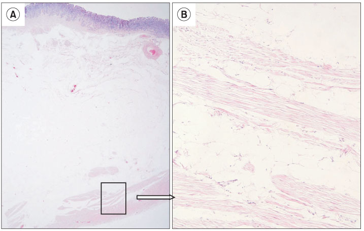
Fig. 4 Photomicrographic findings. Note the mature adipocytes replacing the submucosa and the muscle layer of the stomach (A: H&E, ×10 and B: H&E, ×200)
Reference
-
1. Kim HS, Noh SH, Kim CK. Lipomas of gastrointestinal tract. J Korean Surg Soc. 1989. 36:98–108.
Article2. Ferrozzi F, Tognini G, Bova D, Pavone P. Lipomatous tumors of the stomach: CT findings and differential diagnosis. J Comput Assist Tomogr. 2000. 24:854–858.
Article3. Suárez Moreno RM, Hernández Ramírez DA, Madrazo Navarro M, Salazar Lozano CR, Martínez Gen R. Multiple intestinal lipomatosis. Case report. Cir Cir. 2010. 78:163–165.
Article4. Peabody JW Jr, Zikind J. Lipomatosis of the stomach; a case report and a review of the literature. Ann Surg. 1953. 138:784–790.
Article5. Ventura L, Leocata P, Guadagni S, Ventura T. Multiple gastric lipomas: report of an asymptomatic case found at autopsy. Pathol Int. 1997. 47:575–577.
Article6. Skinner MS, Broadaway RK, Grossman P, Seckinger D. Multiple gastric lipomas. Dig Dis Sci. 1983. 28:1147–1149.
Article7. Deeths TM, Madden PN, Dodds WJ. Multiple lipomas of the stomach and duodenum. Am J Dig Dis. 1975. 20:771–774.
Article8. Devlies F, Hoe LV, Leemans A, Ponette E, Paepe ID. Gastroduodenal lipomatosis. Eur Radiol. 1997. 7:338–340.
Article9. Fawcett NW, Bolton VL, Geever EF. Multiple Lipomas of the Stomach and Duodenum. Ann Surg. 1949. 129:524–527.
Article10. Weinberg T, Feldman M. Lipomas of the gastrointestinal tract. Am J Clin Pathol. 1955. 25:272–281.
Article11. Cabaud PG, Harris LT. Lipomatosis of the ileocecal valve. Ann Surg. 1959. 150:1092–1098.
Article12. Thompson WM, Kende AI, Levy AD. Imaging characteristics of gastric lipomas in 16 adult and pediatric patients. AJR Am J Roentgenol. 2003. 181:981–985.
Article13. Siegal A, Witz M. Gastrointestinal lipoma and malignancies. J Surg Oncol. 1991. 47:170–174.14. The Information Committee of the Korean Gastric Cancer Association. 2005-2006 nationwide gastric submucosal tumor report in Korea. J Korean Gastric Cancer Assoc. 2008. 8:104–111.
