Intrahepatic Splenosis Mimicking Liver Metastasis in a Patient with Gastric Cancer
- Affiliations
-
- 1Department of Surgery, Soonchunhyang University College of Medicine, Bucheon, Korea. gschogs@schbc.ac.kr
- 2Department of Pathology, Soonchunhyang University College of Medicine, Bucheon, Korea.
- 3Department of Radiology, Soonchunhyang University College of Medicine, Bucheon, Korea.
Abstract
- A 54 year old man was referred to our hospital with gastric cancer. The patient had a history of splenectomy and a left nephrectomy as a result of a traffic accident 15 years earlier. The endoscopic findings were advanced gastric cancer at the lower body of the stomach. Abdominal ultrasonography (USG) and magnetic resonance imaging demonstrated a metastatic nodule in the S2 segment of the liver. Eventually, the clinical stage was determined to be cT2cN1cM1 and a radical distal gastrectomy, lateral segmentectomy of the liver were performed. The histopathology findings confirmed the diagnosis of intrahepatic splenosis, omental splenosis. Hepatic splenosis is not rare in patients with a history of splenic trauma or splenectomy. Nevertheless, this is the first report describing a patient with gastric cancer and intrahepatic splenosis that was misinterpreted as a liver metastatic nodule. Intra-operative USG guided fine needle aspiration should be considered to avoid unnecessary liver resections in patients with a suspicious hepatic metastasis.
Keyword
MeSH Terms
Figure
-
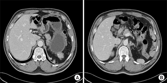
Fig. 1 The axial intravenous contrast enhanced CT scan (the portal venous phase image). (A) Note the wall thickening of the anterior wall of the antrum in the stomach and the lymph node enlargement in the perigastric area. (B) Note the 3.5×1.5 cm sized splenic tissue in the left subdiaphragmatic area with no supplying splenic vessel. CT = computed tomography.
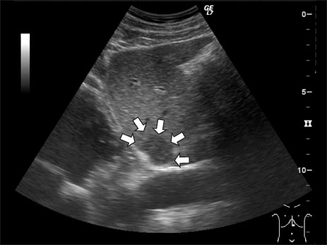
Fig. 2 Ultrasound revealed a 2.5×1.7 cm sized nodular heterogenous hypoechoic lesion with clear margins in segment two of the liver.
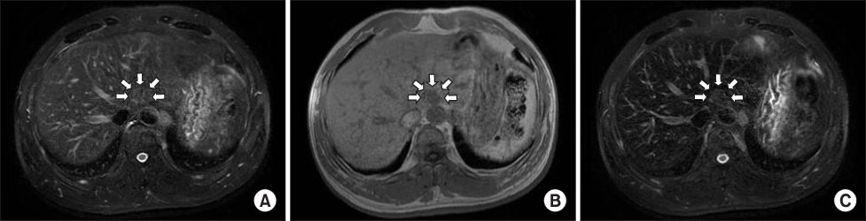
Fig. 3 Axial liver MRI. (A) there was a 2.0×1.5 cm sized nodule in segment two of the liver with slightly high signal intensity on the T2-weighted image and (B) low signal intensity on the T1-weighted image. This nodule still showed high signal intensity on the superparamagnetic iron oxide (SPIO) enhanced T2 weighted image (C). MRI = magnetic resonance imaging.
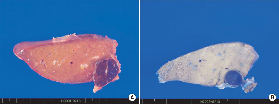
Fig. 4 On the multiple serial sections of the liver, there were two encapsulated red-blue nodules that measured 2.3×1.9 cm (A) and 0.7×0.6 cm (B), respectively. They were located near the liver capsule.
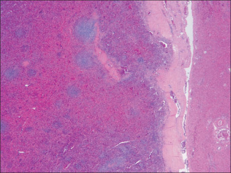
Fig. 5 The pathological examination of this lesion found a full component of red and white pulp of the spleen and this confirmed the diagnosis of splenosis, with no evidence of metastasis (H&E, ×20).
Reference
-
1. Shaw AFB, Shafi A. Traumatic autoplastic transplantation of splenic tissue in man with observations on the late results of splenectomy in six cases. J Pathol. 1937. 45:215–235.
Article2. Fleming CR, Dickson ER, Harrison EG Jr. Splenosis: autotransplantation of splenic tissue. Am J Med. 1976. 61:414–419.
Article3. Marchant LK, Levine MS, Furth EE. Splenic implant in the jejunum: radiographic and pathologic findings. Abdom Imaging. 1995. 20:518–520.
Article4. Bock DB, King BF, Hezmall HP, Oesterling JE. Splenosis presenting as a left renal mass indistinguishable from renal cell carcinoma. J Urol. 1991. 146:152–154.
Article5. De Vuysere S, Van Steenbergen W, Aerts R, Van Hauwaert H, Van Beckevoort D, Van Hoe L. Intrahepatic splenosis: imaging features. Abdom Imaging. 2000. 25:187–189.
Article6. Stein S, Duarte PS, Alavi A, Zhuang H, Alavi JB. Multiple intraabdominal soft-tissue masses in a man awaiting liver transplantation: a case study and discussion. Am J Clin Oncol. 2000. 23:506–508.
Article7. Lee JB, Ryu KW, Song TJ, Suh SO, Kim YC, Koo BH, et al. Hepatic splenosis diagnosed as hepatocellular carcinoma: report of a case. Surg Today. 2002. 32:180–182.
Article8. D'Angelica M, Fong Y, Blumgart LH. Isolated hepatic splenosis: first reported case. HPB Surg. 1998. 11:39–42.9. Kondo M, Okazaki H, Takai K, Nishikawa J, Ohta H, Uekusa T, et al. Intrahepatic splenosis in a patient with chronic hepatitis C. J Gastroenterol. 2004. 39:1013–1015.
Article10. Abu Hilal M, Harb A, Zeidan B, Steadman B, Primrose JN, Pearce NW. Hepatic splenosis mimicking HCC in a patient with hepatitis C liver cirrhosis and mildly raised alpha feto protein; the important role of explorative laparoscopy. World J Surg Oncol. 2009. 7:1.
Article11. Nakajima T, Fujiwara A, Yamaguchi M, Makiyama A, Wakae T, Fujita K, et al. Intrahepatic splenosis with severe iron deposition presenting with atypical magnetic resonance images. Intern Med. 2008. 47:743–746.
Article12. Foroudi F, Ahern V, Peduto A. Splenosis mimicking metastases from breast carcinoma. Clin Oncol (R Coll Radiol). 1999. 11:190–192.
Article13. Shirabe K, Shimada M, Matsumata T, Higashi H, Yakeishi Y, Wakiyama S, et al. Analysis of the prognostic factors for liver metastasis of gastric cancer after hepatic resection: a multi-institutional study of the indications for resection. Hepatogastroenterology. 2003. 50:1560–1563.14. Lin WC, Lee RC, Chiang JH, Wei CJ, Chu LS, Liu RS, et al. MR features of abdominal splenosis. AJR Am J Roentgenol. 2003. 180:493–496.
Article15. Hagman TF, Winer-Muram HT, Meyer CA, Jennings SG. Intrathoracic splenosis: superiority of technetium Tc 99m heat-damaged RBC imaging. Chest. 2001. 120:2097–2098.
- Full Text Links
-
- Actions
-
Cited
- CITED
-
- Close
- Share
-
- Similar articles
-
- Intrahepatic Splenosis Mimicking Hepatocellular Carcinoma: A Case Report
- Splenosis Mimicking Carcinomatosis Peritonei in Advanced Gastric Cancer
- Splenosis Mimicking Recurrence of Renal Cell Carcinoma after Radical Nephrectomy: A Case Report
- Splenosis Mimicking Hepatocellular Carcinoma
- Hepatic Solitary Metastasis of Gastric Cancer: Radiofrequency
