Ann Dermatol.
2016 Dec;28(6):783-784. 10.5021/ad.2016.28.6.783.
A Cutaneous Horn-Like Form of Juvenile Xanthogranuloma
- Affiliations
-
- 1Department of Dermatology, Bucheon St. Mary's Hospital, College of Medicine, The Catholic University of Korea, Bucheon, Korea. beauty4u@catholic.ac.kr
- KMID: 2368138
- DOI: http://doi.org/10.5021/ad.2016.28.6.783
Abstract
- No abstract available.
MeSH Terms
Figure
-
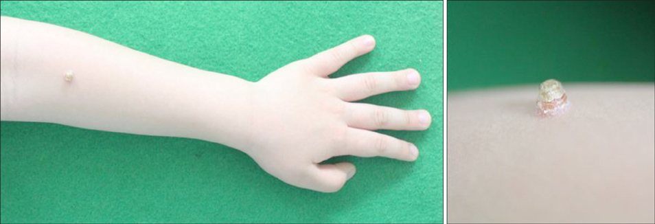
Fig. 1 An asymptomatic, solitary, firm, corn-shaped, yellowish to erythematous nodule with hyperkeratotic cap, present on the left forearm for 2 months.
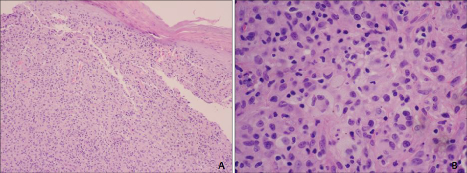
Fig. 2 (A) Dense histiocytic infiltrate in the dermis, including Touton giant cells (H&E, ×100). (B) Foamy cells, lymphocytes, and typical Touton giant cells (H&E, ×400).
Reference
-
1. Hernandez-Martin A, Baselga E, Drolet BA, Esterly NB. Juvenile xanthogranuloma. J Am Acad Dermatol. 1997; 36:355–367. quiz 368-369.
Article2. Caputo R, Grimalt R, Gelmetti C, Cottoni F. Unusual aspects of juvenile xanthogranuloma. J Am Acad Dermatol. 1993; 29:868–870.
Article3. Motegi S, Nagai Y, Amano H, Tamura A, Ishikawa O. An unusual presentation of juvenile xanthogranuloma. Pediatr Dermatol. 2007; 24:576–577.
Article4. Sim JH, Lee ES. Molluscum contagiosum presenting as a cutaneous horn. Ann Dermatol. 2011; 23:262–263.
Article5. Arribas MP, Betlloch I, Soro P, Alfonso R. Giant cutaneous horn on the forearm of a neonate. Juvenile xanthogranuloma. Pediatr Dermatol. 2013; 30:261–262.
