Chonnam Med J.
2017 Jan;53(1):47-55. 10.4068/cmj.2017.53.1.47.
The Clinical Importance of Perforator Preservation in Intracranial Aneurysm Surgery: An Overview with a Review of the Literature
- Affiliations
-
- 1Department of Neurosurgery, Chonnam National University Hospital, Chonnam National University Medical School, Gwangju, Korea. taesun1963@yahoo.co.kr
- KMID: 2367339
- DOI: http://doi.org/10.4068/cmj.2017.53.1.47
Abstract
- Clipping for intracranial aneurysms is done to achieve complete occlusion of the aneurysm without a remnant sac. Despite modern advancements of neurosurgical techniques, morbidity related to the clipping of intracranial aneurysms still exists. Clip occlusion of a parent artery or small hidden perforators commonly leads to permanent neurological deficits, and is a serious and unwanted complication. Thus, preserving blood flow in the branches and perforators of a parent artery is very important for successful surgery without postoperative morbidity and mortality. The aim of this review article is to discuss the consequences of perforator injury and how to avoid this phenomenon in aneurysm surgeries using intraoperative monitoring devices.
Keyword
MeSH Terms
Figure
-
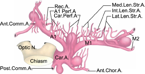
FIG. 1 Schematic illustration of relationships between aneurysms and perforators. The aneurysms involving these perforators arise at four sites: (1) Origin of the anterior choroidal artery, (2) internal carotid bifurcation, (3) middle cerebral artery bifurcation, (4) anterior communicating artery. Courtesy by Rosner et al.33
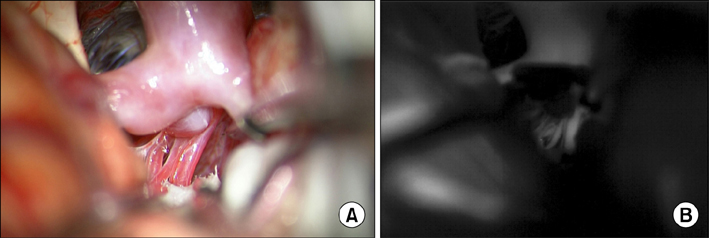
FIG. 2 Representative case of internal carotid artery bifurcation aneruysm. (A) Intraoperative findings of multiple perforators arising behind aneurysm. (B) Indocyanine green video angiography findings showing perforator preservation after aneurysm clipping.
Reference
-
1. Barrow DL, Boyer KL, Joseph GJ. Intraoperative angiography in the management of neurovascular disorders. Neurosurgery. 1992; 30:153–159.
Article2. Siasios I, Kapsalaki EZ, Fountas KN. The role of intraoperative micro-Doppler ultrasound in verifying proper clip placement in intracranial aneurysm surgery. Neuroradiology. 2012; 54:1109–1118.
Article3. Washington CW, Zipfel GJ, Chicoine MR, Derdeyn CP, Rich KM, Moran CJ, et al. Comparing indocyanine green videoangiography to the gold standard of intraoperative digital subtraction angiography used in aneurysm surgery. J Neurosurg. 2013; 118:420–427.
Article4. de Oliveira JG, Beck J, Seifert V, Teixeira MJ, Raabe A. Assessment of flow in perforating arteries during intracranial aneurysm surgery using intraoperative near-infrared indocyanine green videoangiography. Neurosurgery. 2007; 61:3 Suppl. 63–72. discussion 72-3.
Article5. Raabe A, Beck J, Gerlach R, Zimmermann M, Seifert V. Near-infrared indocyanine green video angiography: a new method for intraoperative assessment of vascular flow. Neurosurgery. 2003; 52:132–139. discussion 139.
Article6. Fischer G, Stadie A, Oertel JM. Near-infrared indocyanine green videoangiography versus microvascular Doppler sonography in aneurysm surgery. Acta Neurochir (Wien). 2010; 152:1519–1525.
Article7. Kalavakonda C, Sekhar LN, Ramachandran P, Hechl P. Endoscope-assisted microsurgery for intracranial aneurysms. Neurosurgery. 2002; 51:1119–1126. discussion 1126-7.
Article8. Kinouchi H, Yanagisawa T, Suzuki A, Ohta T, Hirano Y, Sugawara T, et al. Simultaneous microscopic and endoscopic monitoring during surgery for internal carotid artery aneurysms. J Neurosurg. 2004; 101:989–995.
Article9. Taniguchi M, Takimoto H, Yoshimine T, Shimada N, Miyao Y, Hirata M, et al. Application of a rigid endoscope to the microsurgical management of 54 cerebral aneurysms: results in 48 patients. J Neurosurg. 1999; 91:231–237.
Article10. Kato Y, Sano H, Nagahisa S, Iwata S, Yoshida K, Yamamoto K, et al. Endoscope-assisted microsurgery for cerebral aneurysms. Minim Invasive Neurosurg. 2000; 43:91–97.
Article11. van Lindert E, Perneczky A, Fries G, Pierangeli E. The supraorbital keyhole approach to supratentorial aneurysms: concept and technique. Surg Neurol. 1998; 49:481–489. discussion 489-90.
Article12. Martin NA, Bentson J, Viñuela F, Hieshima G, Reicher M, Black K, et al. Intraoperative digital subtraction angiography and the surgical treatment of intracranial aneurysms and vascular malformations. J Neurosurg. 1990; 73:526–533.
Article13. Mielke D, Malinova V, Rohde V. Comparison of intraoperative microscopic and endoscopic ICG angiography in aneurysm surgery. Neurosurgery. 2014; 10:Suppl 3. 418–425. discussion 425.
Article14. Gruber A, Dorfer C, Standhardt H, Bavinzski G, Knosp E. Prospective comparison of intraoperative vascular monitoring technologies during cerebral aneurysm surgery. Neurosurgery. 2011; 68:657–673. discussion 673.
Article15. Pritz MB. Cerebral aneurysm classification based on angioarchitecture. J Stroke Cerebrovasc Dis. 2011; 20:162–167.
Article16. Pritz MB. Perforator and secondary branch origin in relation to the neck of saccular, cerebral bifurcation aneurysms. World Neurosurg. 2014; 82:726–732.
Article17. Caruso G, Vincentelli F, Giudicelli G, Grisoli F, Xu T, Gouaze A. Perforating branches of the basilar bifurcation. J Neurosurg. 1990; 73:259–265.
Article18. Tulleken CA, Luiten ML. The basilar artery bifurcation: microscopical anatomy. Acta Neurochir (Wien). 1987; 85:50–55.
Article19. Pedroza A, Dujovny M, Ausman JI, Diaz FG, Cabezudo Artero J, et al. Microvascular anatomy of the interpeduncular fossa. J Neurosurg. 1986; 64:484–493.
Article20. Da Pian R, Pasqualin A, Scienza R. Direct microsurgical approach to aneurysms of the internal carotid bifurcation. Surg Neurol. 1980; 13:27–37.21. Kyoshima K, Kobayashi S, Nitta J, Osawa M, Shigeta H, Nakagawa F. Clinical analysis of internal carotid artery aneurysms with reference to classification and clipping techniques. Acta Neurochir (Wien). 1998; 140:933–942.
Article22. Laranjeira M, Sadasivan B, Ausman JI. Direct surgery for carotid bifurcation artery aneurysms. Surg Neurol. 1990; 34:250–254.
Article23. Miyazawa N, Nukui H, Horikoshi T, Yagishita T, Sugita M, Kanemaru K. Surgical management of aneurysms of the bifurcation of the internal carotid artery. Clin Neurol Neurosurg. 2002; 104:103–114.
Article24. van Rooij WJ, Sluzewski M, Beute GN. Internal carotid bifurcation aneurysms: frequency, angiographic anatomy and results of coiling in 50 aneurysms. Neuroradiology. 2008; 50:583–587.
Article25. Lehecka M, Dashti R, Romani R, Celik O, Navratil O, Kivipelto L, et al. Microneurosurgical management of internal carotid artery bifurcation aneurysms. Surg Neurol. 2009; 71:649–667.
Article26. Dashti R, Hernesniemi J, Lehto H, Niemelä M, Lehecka M, Rinne J, et al. Microneurosurgical management of proximal anterior cerebral artery aneurysms. Surg Neurol. 2007; 68:366–377.
Article27. Dashti R, Rinne J, Hernesniemi J, Niemelä M, Kivipelto L, Lehecka M, et al. Microneurosurgical management of proximal middle cerebral artery aneurysms. Surg Neurol. 2007; 67:6–14.
Article28. Gibo H, Lenkey C, Rhoton AL Jr. Microsurgical anatomy of the supraclinoid portion of the internal carotid artery. J Neurosurg. 1981; 55:560–574.
Article29. Marinković S, Gibo H, Milisavljević M. The surgical anatomy of the relationships between the perforating and the leptomeningeal arteries. Neurosurgery. 1996; 39:72–83.
Article30. Marinković SV, Milisavljević MM, Marinković ZD. The perforating branches of the internal carotid artery: the microsurgical anatomy of their extracerebral segments. Neurosurgery. 1990; 26:472–478. discussion 478-9.31. Türe U, Yaşargil MG, Al-Mefty O, Yaşargil DC. Arteries of the insula. J Neurosurg. 2000; 92:676–687.
Article32. Rinne J, Hernesniemi J, Niskanen M, Vapalahti M. Analysis of 561 patients with 690 middle cerebral artery aneurysms: anatomic and clinical features as correlated to management outcome. Neurosurgery. 1996; 38:2–11.
Article33. Rosner SS, Rhoton AL Jr, Ono M, Barry M. Microsurgical anatomy of the anterior perforating arteries. J Neurosurg. 1984; 61:468–485.
Article34. Perlmutter D, Rhoton AL Jr. Microsurgical anatomy of anterior cerebral anterior communicating recurrent artery complex. Surg Forum. 1976; 27:464–465.35. Handa J, Nakasu Y, Matsuda M, Kyoshima K. Aneurysms of the proximal anterior cerebral artery. Surg Neurol. 1984; 22:486–490.
Article36. Hino A, Fujimoto M, Iwamoto Y, Oka H, Echigo T. Surgery of proximal anterior cerebral artery aneurysms. Acta Neurochir (Wien). 2002; 144:1291–1296. discussion 1296.37. Suzuki M, Onuma T, Sakurai Y, Mizoi K, Ogawa A, Yoshimoto T. Aneurysms arising from the proximal (A1) segment of the anterior cerebral artery. A study of 38 cases. J Neurosurg. 1992; 76:455–458.
Article38. Wakabayashi T, Tamaki N, Yamashita H, Saya H, Suyama T, Matsumoto S. Angiographic classification of aneurysms of the horizontal segment of the anterior cerebral artery. Surg Neurol. 1985; 24:31–34.
Article39. Wanibuchi M, Kurokawa Y, Ishiguro M, Fujishige M, Inaba K. Characteristics of aneurysms arising from the horizontal portion of the anterior cerebral artery. Surg Neurol. 2001; 55:148–154. discussion 154-5.
Article40. Lee JM, Joo SP, Kim TS, Go EJ, Choi HY, Seo BR. Surgical management of anterior cerebral artery aneurysms of the proximal (A1) segment. World Neurosurg. 2010; 74:478–482.
Article41. Evans AL, Corkill RA, Wenderoth JD. Ruptured fusiform aneurysm of fenestrated A1 segment of the anterior cerebral artery. Case report and review of the literature. Neuroradiology. 2006; 48:196–199.
Article42. Hirao J, Okamoto H, Watanabe T, Asano S, Teraoka A. Dissecting aneurysms at the A1 segment of the anterior cerebral artery--two case reports. Neurol Med Chir (Tokyo). 2001; 41:271–278.
Article43. Kashimura H, Mase T, Ogasawara K, Ogawa A, Endo H. Trapping and vascular reconstruction for ruptured fusiform aneurysm in the proximal A1 segment of the anterior cerebral artery. Neurol Med Chir (Tokyo). 2006; 46:340–343.
Article44. Nomura M, Kida S, Kita D, Higashi R, Hasegawa M, Matsui O, et al. Fusiform aneurysm of the proximal anterior cerebral artery (A1). Acta Neurochir (Wien). 2000; 142:1163–1164.
Article45. Dunker RO, Harris AB. Surgical anatomy of the proximal anterior cerebral artery. J Neurosurg. 1976; 44:359–367.
Article46. Ostrowski AZ, Webster JE, Gurdjian ES. The proximal anterior cerebral artery: an anatomic study. Arch Neurol. 1960; 3:661–664.
Article47. Gomes F, Dujovny M, Umansky F, Ausman JI, Diaz FG, Ray WJ, et al. Microsurgical anatomy of the recurrent artery of Heubner. J Neurosurg. 1984; 60:130–139.
Article48. Tulleken CA. A study of the anatomy of the anterior communicating artery with the aid of the operating microscope. Clin Neurol Neurosurg. 1978; 80:169–173.
Article49. Drake CG, Vanderlinden RG, Amacher AL. Carotid-choroidal aneurysms. J Neurosurg. 1968; 29:32–36.
Article50. Friedman JA, Pichelmann MA, Piepgras DG, Atkinson JL, Maher CO, Meyer FB, et al. Ischemic complications of surgery for anterior choroidal artery aneurysms. J Neurosurg. 2001; 94:565–572.
Article51. Yasargil MG, Yonas H, Gasser JC. Anterior choroidal artery aneurysms: their anatomy and surgical significance. Surg Neurol. 1978; 9:129–138.52. Erdem A, Yaşargil G, Roth P. Microsurgical anatomy of the hippocampal arteries. J Neurosurg. 1993; 79:256–265.
Article53. Rhoton AL Jr, Fujii K, Fradd B. Microsurgical anatomy of the anterior choroidal artery. Surg Neurol. 1979; 12:171–187.54. Fujii K, Lenkey C, Rhoton AL Jr. Microsurgical anatomy of the choroidal arteries. Fourth ventricle and cerebellopontine angles. J Neurosurg. 1980; 52:504–524.
Article55. Marinković S, Gibo H, Brigante L, Nikodijević I, Petrović P. The surgical anatomy of the perforating branches of the anterior choroidal artery. Surg Neurol. 1999; 52:30–36.
Article56. Saeki N, Rhoton AL Jr. Microsurgical anatomy of the upper basilar artery and the posterior circle of Willis. J Neurosurg. 1977; 46:563–578.
Article57. Yoshioka H, Kinouchi H. The roles of endoscope in aneurysmal surgery. Neurol Med Chir (Tokyo). 2015; 55:469–478.
Article58. Apuzzo ML, Heifetz MD, Weiss MH, Kurze T. Neurosurgical endoscopy using the side-viewing telescope. J Neurosurg. 1977; 46:398–400.
Article59. Cohen AR, Perneczky A, Rodziewicz GS, Gingold SI. Endoscope-assisted craniotomy: approach to the rostral brain stem. Neurosurgery. 1995; 36:1128–1129. discussion 1129-30.60. Fischer G, Oertel J, Perneczky A. Endoscopy in aneurysm surgery. Neurosurgery. 2012; 70:2 Suppl Operative. 184–190. discussion 190-1.
Article61. Fischer J, Mustafa H. Endoscopic-guided clipping of cerebral aneurysms. Br J Neurosurg. 1994; 8:559–565.
Article62. Galzio RJ, Di Cola F, Raysi Dehcordi S, Ricci A, De Paulis D. Endoscope-assisted microneurosurgery for intracranial aneurysms. Front Neurol. 2013; 4:201.
Article63. Profeta G, De Falco R, Ambrosio G, Profeta L. Endoscope-assisted microneurosurgery for anterior circulation aneurysms using the angle-type rigid endoscope over a 3-year period. Childs Nerv Syst. 2004; 20:811–815.
Article64. Wang E, Yong NP, Ng I. Endoscopic assisted microneurosurgery for cerebral aneurysms. J Clin Neurosci. 2003; 10:174–176.65. Fries G, Perneczky A. Endoscope-assisted brain surgery: part 2--analysis of 380 procedures. Neurosurgery. 1998; 42:226–231. discussion 231-2.
Article66. Perneczky A, Fries G. Endoscope-assisted brain surgery: part 1--evolution, basic concept, and current technique. Neurosurgery. 1998; 42:219–224. discussion 224-5.
Article67. Kuroda K, Kinouchi H, Kanemaru K, Nishiyama Y, Ogiwara M, Yoshioka H, et al. Intra-arterial injection fluorescein videoangiography in aneurysm surgery. Neurosurgery. 2013; 72:2 Suppl Operative. ons141–ons150. discussion ons150.
Article68. Raabe A, Nakaji P, Beck J, Kim LJ, Hsu FP, Kamerman JD, et al. Prospective evaluation of surgical microscope-integrated intraoperative near-infrared indocyanine green videoangiography during aneurysm surgery. J Neurosurg. 2005; 103:982–989.
Article69. Yoshioka H, Kinouchi H, Nishiyama Y, Kanemaru K, Yagi T, Hanihara M, et al. Advantage of microscope integrated for both indocyanine green and fluorescein videoangiography on aneurysmal surgery: case report. Neurol Med Chir (Tokyo). 2014; 54:192–195.
Article70. Wrobel CJ, Meltzer H, Lamond R, Alksne JF. Intraoperative assessment of aneurysm clip placement by intravenous fluorescein angiography. Neurosurgery. 1994; 35:970–973. discussion 973.
Article71. Suzuki K, Kodama N, Sasaki T, Matsumoto M, Ichikawa T, Munakata R, et al. Confirmation of blood flow in perforating arteries using fluorescein cerebral angiography during aneurysm surgery. J Neurosurg. 2007; 107:68–73.
Article72. Gilsbach JM, Harders AG. Microvascular and transcranial Doppler sonographic evaluation of cerebral aneurysm flow pattern. Neurol Res. 1989; 11:41–48.
Article73. Gilsbach JM, Hassler WE. Intraoperative Doppler and real time sonography in neurosurgery. Neurosurg Rev. 1984; 7:199–208.
Article74. Marchese E, Albanese A, Denaro L, Vignati A, Fernandez E, Maira G. Intraoperative microvascular Doppler in intracranial aneurysm surgery. Surg Neurol. 2005; 63:336–342. discussion 342.
Article75. Cui H, Wang Y, Yin Y, Wan J, Fei Z, Gao W, et al. Role of intraoperative microvascular Doppler in the microsurgical management of intracranial aneurysms. J Clin Ultrasound. 2011; 39:27–31.
Article76. Ishizaki T, Endo O, Fujii K, Matsudaira T, Okada T, Kobayashi N, et al. Usefulness and problems of intraoperative monitoring for unruptured aneurysm surgery with the motor evoked potential. No Shinkei Geka. 2016; 44:283–293.
- Full Text Links
-
- Actions
-
Cited
- CITED
-
- Close
- Share
-
- Similar articles
-
- Intracranial Aneurysm Associated with Moyamoya Disease
- Ruptured Intracranial Dermoid Cyst Associated with Rupture of Cerebral Aneurysm
- A Case of Intracranial Mycotic Aneurysm due to Aspergillus species
- Surgical Principles and Tactics for Successful Clipping of Intracranial Aneurysms
- Ruptured Intracranial Aneurysm in a 45-day-old Infant
