Retreatment of failed regenerative endodontic of orthodontically treated immature permanent maxillary central incisor: a case report
- Affiliations
-
- 1Security Force Hospital, Dental Department, Riyadh, Saudi Arabia.
- 2Department of Restorative Dental Science, Division of Endodontics, King Saudi University, College of Dentistry, Riyadh, Saudi Arabia. snazhan@ksu.edu.sa
- KMID: 2367324
- DOI: http://doi.org/10.5395/rde.2017.42.1.65
Abstract
- A revascularization procedure was shown to be the best alternative therapy for immature teeth with necrotic pulp and apical infection. A 12 year old female with a history of trauma to her upper central incisor and a sinus tract was referred for endodontic treatment. She was an active orthodontic patient and had undergone regenerative endodontic treatment for the past 2 years. Clinical examination revealed no response to sensibility, percussion, and palpation tests. The preoperative radiograph showed an open apex and apical rarefaction. The case was diagnosed as previously treated tooth with asymptomatic apical periodontitis. Regenerative endodontic retreatment was performed, and the case was followed for 3 years. Clinical, radiographic, and cone-beam computed tomography follow-up examination revealed an asymptomatic tooth, with evidence of periapical healing and root maturation.
Keyword
MeSH Terms
Figure
-

Figure 1 (a) Diagnostic radiograph of the maxillary right central incisor (tooth #11) demonstrated incomplete root formation with diffuse periapical radiolucency and poor root canal filling; (b and c) Preoperative clinical photograph illustrates orthodontic treatment and the sinus tract related to tooth #11; (d) Periapical radiograph during orthodontic examination and before regenerative endodontic treatment that shows an open apex with apical rarefaction.
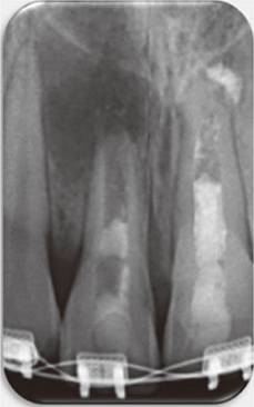
Figure 2 Periapical radiograph after placement of double antibiotic paste (DAP).
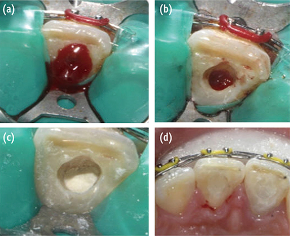
Figure 3 Second appointment. (a) Bleeding created by overinstrumentation; (b) Bleeding stopped 3 mm from CEJ; (c) Collagen membrane placed on top of the blood clot and placement of white MTA; (d) Access cavity restored with composite resin restoration. CEJ, cementoenamel junction; MTA, mineral trioxide aggregate.
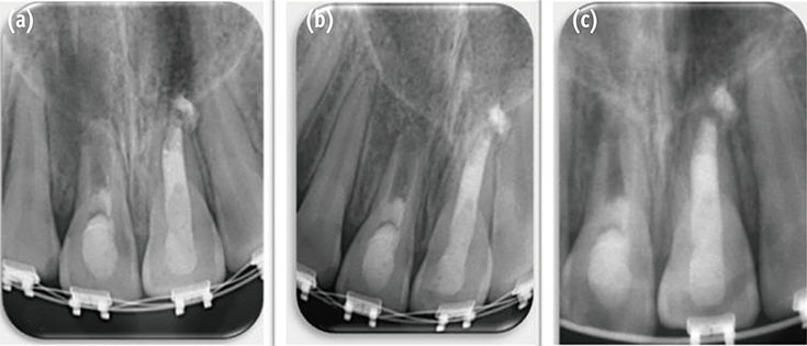
Figure 4 Follow-up radiographs of tooth #11 at (a) 3 months; (b) 6 months; (c) 12 months.
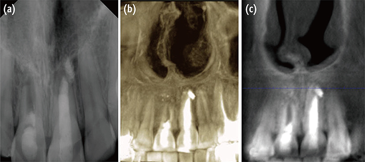
Figure 5 Three year follow-up confirming the healing process. (a) Conventional radiograph; (b) CBCT three-dimensional reconstruction; (c) CBCT buccal view. CBCT, cone beam computed tomography.
Cited by 1 articles
-
Clinical and radiographic outcomes of regenerative endodontic treatment performed by endodontic postgraduate students: a retrospective study
Hadi Rajeh Alfahadi, Saad Al-Nazhan, Fawaz Hamad Alkazman, Nassr Al-Maflehi, Nada Al-Nazhan
Restor Dent Endod. 2022;47(2):e24. doi: 10.5395/rde.2022.47.e24.
Reference
-
1. Andreasen JO, Ravn JJ. Epidemiology of traumatic dental injuries to primary and permanent teeth in a Danish population sample. Int J Oral Surg. 1972; 1:235–239.
Article2. Morse DR, O'Larnic J, Yesilsoy C. Apexification: review of the literature. Quintessence Int. 1990; 21:589–598.3. Pace R, Giuliani V, Pini Prato L, Baccetti T, Pagavino G. Apical plug technique using mineral trioxide aggregate: results from a case series. Int Endod J. 2007; 40:478–484.
Article4. Chala S, Abouqal R, Rida S. Apexification of immature teeth with calcium hydroxide or mineral trioxide aggregate: systematic review and meta-analysis. Oral Surg Oral Med Oral Pathol Oral Radiol Endod. 2011; 112:e36–e42.
Article5. Sheehy EC, Roberts GJ. Use of calcium hydroxide for apical barrier formation and healing in non-vital immature permanent teeth: a review. Br Dent J. 1997; 183:241–246.
Article6. Simon S, Rillard F, Berdal A, Machtou P. The use of mineral trioxide aggregate in one visit apexification treatment: a prospective study. Int Endod J. 2007; 40:186–197.
Article7. Andreason JO, Farik B, Munksgaard EC. Long-term calcium hydroxide as a root canal dressing may increase risk of root fracture. Dent Traumatol. 2002; 18:134–137.
Article8. Andreasen JO, Munksgaard EC, Bakland LK. Comparison of fracture resistance in root canals of immature sheep teeth after filling with calcium hydroxide or MTA. Dent Traumatol. 2006; 22:154–156.
Article9. Nagy MM, Tawfik HE, Hashem AA, Abu-Seida AM. Regenerative potential of immature permanent teeth with necrotic pulps after different regenerative protocols. J Endod. 2014; 40:192–198.
Article10. Petrino JA, Boda KK, Shambarger S, Bowles WR, McClanahan SB. Challenges in regenerative endodontics: a case series. J Endod. 2010; 36:536–541.
Article11. Cehreli ZC, Isbitiren B, Sara S, Erbas G. Regenerative endodontic treatment (revascularization) of immature necrotic molars medicated with calcium hydroxide: a case series. J Endod. 2011; 37:1327–1330.
Article12. Al-Ghamdi NS, Al-Nazhan S. Pulp revascularization of immature maxillary first premolar. J Conserv Dent. 2015; 18:496–499.
Article13. Garcia-Godoy F, Murray PE. Recommendations for using regenerative endodontic procedures in permanent immature traumatized teeth. Dent Traumatol. 2012; 28:33–41.
Article14. Jeeruphan T, Jantarat J, Yanpiset K, Suwannapan , Khewsawai P, Hargreaves KM. Mahidol study 1: comparison of radiographic and survival outcomes of immature teeth treated with either regenerative endodontic or apexification methods: a retrospective study. J Endod. 2012; 38:1330–1336.
Article15. Miltiadous ME, Floratos SG. Regenerative endodontic treatment as a retreatment option for a tooth with open apex - a case report. Braz Dent J. 2015; 26:552–556.
Article16. Andreasen JO, Bakland LK, Flores MT, Andreasen FM, Andersson L. Traumatic dental injuries: a manual. 3rd ed. West Sussex: Wiley-Blackwell;2011. p. 12p. 29p. 35p. 63–72.17. Iwaya SI, Ikawa M, Kubota M. Revascularization of an immature permanent tooth with apical periodontitis and sinus tract. Dent Traumatol. 2001; 17:185–187.
Article18. Chen MY, Chen KL, Chen CA, Tayebaty F, Rosenberg PA, Lin LM. Responses of immature permanent teeth with infected necrotic pulp tissue and apical periodontitis/abscess to revascularization procedures. Int Endod J. 2012; 45:294–305.
Article19. Nosrat A, Homayounfar N, Oloomi K. Drawbacks and unfavorable outcomes of regenerative endodontic treatments of necrotic immature teeth: a literature review and report of a case. J Endod. 2012; 38:1428–1434.
Article20. Bose R, Nummikoski P, Hargreaves K. A retrospective evaluation of radiographic outcomes in immature teeth with necrotic root canal systems treated with regenerative endodontic procedures. J Endod. 2009; 35:1343–1349.
Article21. Wang Y, Zhu X, Zhang C. Pulp revascularization on permanent teeth with open apices in a middle-aged patient. J Endod. 2015; 41:1571–1575.
Article22. Jadhav G, Shah N, Logani A. Revascularization with and without platelet-rich plasma in nonvital, immature, anterior teeth: a pilot clinical study. J Endod. 2012; 38:1581–1587.
Article23. Bezgin T, Yilmaz AD, Celik BN, Sönmez H. Concentrated platelet-rich plasma used in root canal revascularization: 2 case reports. Int Endod J. 2014; 47:41–49.
Article24. Kim JH, Kim Y, Shin SJ, Park JW, Jung IY. Tooth discoloration of immature permanent incisor associated with triple antibiotic therapy: a case report. J Endod. 2010; 36:1086–1091.
Article25. Lenzi R, Trope M. Revitalization procedures in two traumatized incisors with different biological outcomes. J Endod. 2012; 38:411–414.
Article26. Thibodeau B, Teixeira F, Yamauchi M, Caplan DJ, Trope M. Pulp revascularization of immature dog teeth with apical periodontitis. J Endod. 2007; 33:680–689.
Article27. Lin L, Shovlin F, Skribner J, Langeland K. Pulp biopsies from the teeth associated with periapical radiolucency. J Endod. 1984; 10:436–448.
Article28. Shi S, Gronthos S. Perivascular niche of postnatal mesenchymal stem cells in human bone marrow and dental pulp. J Bone Miner Res. 2003; 18:696–704.
Article29. Nosrat A, Li KL, Vir K, Hicks ML, Fouad AF. Is pulp regeneration necessary for root maturation? J Endod. 2013; 39:1291–1295.
Article30. Lin LM, Rosenberg PA. Repair and regeneration in endodontics. Int Endod J. 2011; 44:889–906.
Article31. Mavragani M, Bøe OE, Wisth PJ, Selvig KA. Changes in root length during orthodontic treatment: advantages for immature teeth. Eur J Orthod. 2002; 24:91–97.
Article32. de Souza RS, Gandini LG Jr, de Souza V, Holland R, Dezan E Jr. Influence of orthodontic dental movement on the healing process of teeth with periapical lesions. J Endod. 2006; 32:115–119.
Article33. Beck VJ, Stacknik S, Chandler NP, Farella M. Orthodontic tooth movement of traumatised or root-canal-treated teeth: a clinical review. N Z Dent J. 2013; 109:6–11.34. Drysdale C, Gibbs SL, Ford TR. Orthodontic management of root-filled teeth. Br J Orthod. 1996; 23:255–260.
Article35. Malmgren O, Malmgren B. Orthodontic management of the traumatised dentition. In : Andreasen JO, Andreasen FM, Andersson L, editors. Textbook and color atlas of traumatic injuries to the teeth. 4th ed. Oxford: Blackwell;2007. p. 669–715.36. Nagata JY, Gomes BP, Rocha Lima TF, Murakami LS, de Faria DE, Campos GR, de Souza-Filho FJ, Soares Ade J. Traumatized immature teeth treated with 2 protocols of pulp revascularization. J Endod. 2014; 40:606–612.
Article37. American Association of Endodontists: Clinical resources, regenerative endodontics. AAE clinical considerations for a regenerative procedure. updated Oct 9, 2016. Available from: http://www.aae.org/uploadedfiles/publications_and_research/research/currentregenerativeendodonticconsiderations.pdf.38. Ruparel NB, Teixeira FB, Ferraz CC, Diogenes A. Direct effect of intracanal medicaments on survival of stem cells of the apical papilla. J Endod. 2012; 38:1372–1375.
Article39. Chuensombat S, Khemaleelakul S, Chattipakorn S, Srisuwan T. Cytotoxic effects and antibacterial efficacy of a 3-antibiotic combination: an in vitro study. J Endod. 2013; 39:813–819.
Article40. Yassen GH, Chu TM, Eckert G, Platt JA. Effect of medicaments used in endodontic regeneration technique on the chemical structure of human immature radicular dentin: an in vitro study. J Endod. 2013; 39:269–273.
Article41. Maruyama H, Aoki A, Sasaki KM, Takasaki AA, Iwasaki K, Ichinose S, Oda S, Ishikawa I, Izumi Y. The effect of chemical and/or mechanical conditioning on the Er:YAG laser-treated root cementum: analysis of surface morphology and periodontal ligament fibroblast attachment. Lasers Surg Med. 2008; 40:211–222.
Article42. Gomes BP, Vianna ME, Zaia AA, Almeida JF, Souza-Filho FJ, Ferraz CC. Chlorhexidine in endodontics. Braz Dent J. 2013; 24:89–102.
Article43. Trevino EG, Patwardhan AN, Henry MA, Perry G, Dybdal-Hargreaves N, Hargreaves KM, Diogenes A. Effect of irrigants on the survival of human stem cells of the apical papilla in a platelet-rich plasma scaffold in human root tips. J Endod. 2011; 37:1109–1115.
Article44. Forghani M, Parisay I, Maghsoudlou A. Apexogenesis and revascularization treatment procedures for two traumatized immature permanent maxillary incisors: a case report. Restor Dent Endod. 2013; 38:178–181.
Article45. Asgary S, Eghbal MJ, Parirokh M, Ghoddusi J. Effect of two storage solutions on surface topography of two root-end fillings. Aust Endod J. 2009; 35:147–152.
Article46. Sarkar NK, Caicedo R, Ritwik P, Moiseyeva R, Kawashima I. Physicochemical basis of the biologic properties of mineral trioxide aggregate. J Endod. 2005; 31:97–100.
Article
- Full Text Links
-
- Actions
-
Cited
- CITED
-
- Close
- Share
-
- Similar articles
-
- Apexogenesis and revascularization treatment procedures for two traumatized immature permanent maxillary incisors: a case report
- Outcome of Regenerative Endodontic Treatment for an Avulsed Immature Permanent Tooth: A Case Report
- Multidisciplinary management of a fused maxillary central incisor moved through the midpalatal suture: A case report
- Regenerative Endodontic Procedure in Korean Children and Adolescents: A Case Report
- Clinical and radiographic outcomes of regenerative endodontic treatment performed by endodontic postgraduate students: a retrospective study
