Primary Intracranial Malignant Melanoma with Extracranial Metastasis
- Affiliations
-
- 1Department of Neurosurgery, Tokyo Women’s Medical University Medical Center East, Tokyo, Japan. k2bumps@gmail.com
- KMID: 2365594
- DOI: http://doi.org/10.3340/jkns.2015.0506.007
Abstract
- We report a case of primary intracranial malignant melanoma (PIMM) with extracranial metastases. The patient was an 82-year-old woman diagnosed with PIMM under the left cerebellar tentorium. We performed a tumor resection followed by gamma knife surgery. An magnetic resonance imaging at 11 months after surgery showed a local intracranial recurrence. At 12 months, vertebral metastasis was suspected, and 2-[fluorine-18]-fluoro-2-deoxy-D-glucose positron emission tomography/computed tomography (FDG-PET/CT) showed multiple extracranial metastases. She died at 13 months after surgery. Although extracranial metastases of PIMM are extremely rare, we should carefully follow up extracranial metastases together with intracranial ones, especially by FDG-PET/CT, even at an early asymptomatic stage.
MeSH Terms
Figure
-
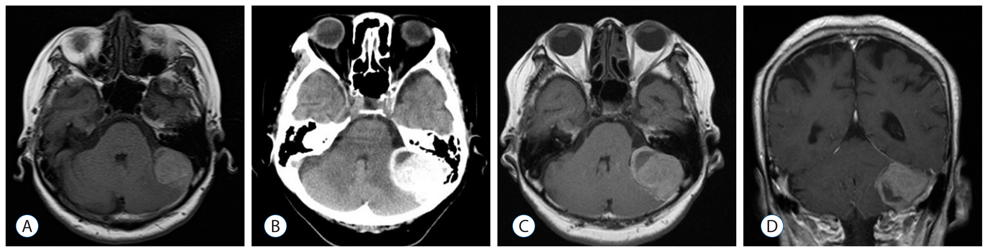
Fig. 1 Axial T1-weighted MRI (A) shows an extra-axial mass lesion under the left cerebellar tentorium one month before admission. Axial CT (B), T1-weighted MRI (C) and Coronal Gd-enhanced T1-weighted MRI (D) on admission show the enlarged mass lesion with niveau formation at one month later. MRI: magnetic resonance imaging, CT: computed tomography.
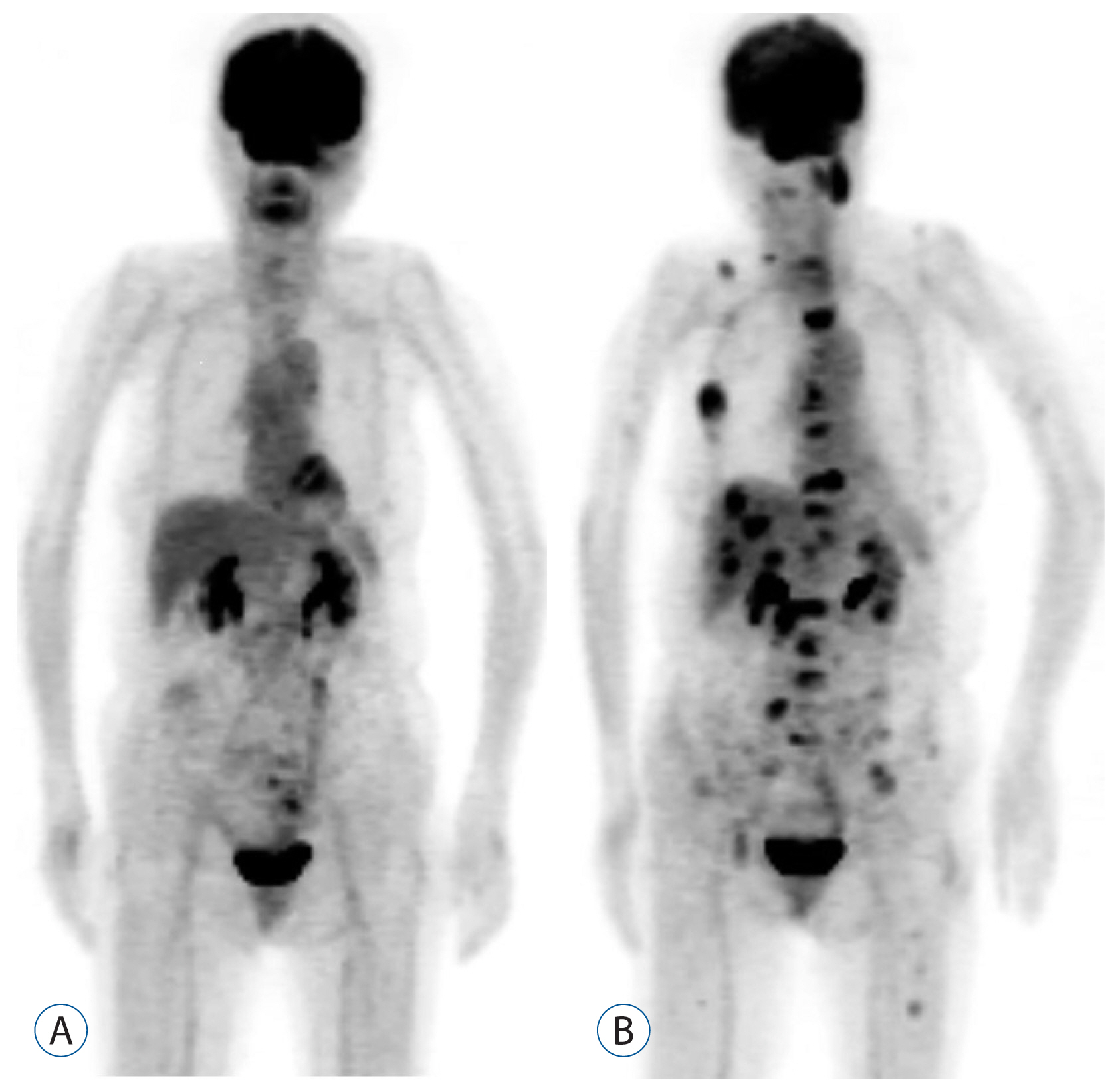
Fig. 2 Whole body FDG-PET/CT (A) on admission shows no malignant lesions such as extracranial malignant melanoma. FDG-PET/CT (B) at 12 months after the surgery shows FDG uptake in multiple metastatic lesions. FDG-PET/CT: 2-[fluorine-18]-fluoro-2-deoxy-D-glucose positron emission tomography/computed tomography.

Fig. 3 Intraoperative image (A) shows the black colored tumor on the left cerebellar tentorium. Photomicrographs. The tumor cells arranged in fascicles intersperse with melanin-containing cells (B, H&E, ×200). The tumor cells are positive for Melan-A (C, ×400). The MIB-1 (Ki-67) staining index is approximately 8% (D, ×400).
Reference
-
References
1. Brat DJ, Giannini C, Scheithauer BW, Burger PC. Primary melanocytic neoplasms of the central nervous systems. Am J Surg Pathol. 23:745–754. 1999.2. Brat DJ, Perry A. Melanocytic lesions. Louis DN, Ohgaki H, Wiestler OD, Cavenee WK, editors. WHO classification of tumours of the central nervous system. 4th ed. Lyon: IARC;2007. p. 181–183.3. Dinesh SM, Suneetha B, Sen A. A rare case of primary malignant melanoma of clivus with extensive skeletal metastasis demonstrated on 18F-FDG PET/CT. Indian J Nucl Med. 28:234–236. 2013.
Article4. Glasauer FE, Yuan RH. Intracranial tumors with extracranial metastases. Case report and review of the literature. J Neurosurg. 20:474–493. 1963.5. Crasto Greco S, Soffietti R, Bradac GB, Boldorini R. Primitive cerebral melanoma: case report and review of the literature. Surg Neurol. 55:163–168. discussion 168. 2001.
Article6. Hayward RD. Malignant melanoma and the central nervous system: a guide for classification based on clinical findings. J Neurol Neurosurg Psychiatr. 39:526–530. 1976.
Article7. Hoffman HJ, Duffner PK. Extraneural metastases of central nervous system tumors. Cancer. 56(7 Suppl):1778–1782. 1985.
Article8. Koskivuo IO, Seppänen MP, Suominen EA, Minn HR. Whole body positron emission tomography in follow-up of high risk melanoma. Acta Oncol. 46:685–690. 2007.
Article9. Kim MS, Yoon DH, Shin DA. Primary spinal cord melanoma. J Korean Neurosurg Soc. 48:157–161. 2010.
Article10. Larcos G, Maisey MN. FDG-PET screening for cerebral metastases in patients with suspected malignancy. Nucl Med Commun. 17:197–198. 1996.
Article11. Lun M, Lok E, Gautam S, Wu E, Wong ET. The natural history of extracranial metastasis from glioblastoma multiforme. J Neurooncol. 105:261–273. 2011.
Article12. Nakagawa H, Hayakawa T, Niiyama K, Nii Y, Yoshimine T, Mori S. Long-term survival after removal of primary intracranial malignant melanoma. Case report. Acta Neurochir (Wien). 101:84–88. 1989.
Article13. Rodriguez y Baena R, Gaetani P, Danova M, Bosi F, Zappoli F. Primary solitary intracranial melanoma: case report and review of literature. Surg Neurol. 38:26–37. 1992.
Article14. Shah I, Imran M, Akram R, Rafat S, Zia K, Emaduddin M. Primary intracranial malignant melanoma. J Coll Physicians Surg Pak. 23:157–159. 2013.15. Wadasadawala T, Trivedi S, Gupta T, Epari S, Jalali R. The diagnostic dilemma of primary central nervous system melanoma. J Clin Neurosci. 17:1014–1017. 2010.
Article16. Wang J, Guo ZZ, Wang YJ, Zhang SG, Xing DG. Microsurgery for the treatment of primary malignant intracranial melanoma: a surgical series and literature review. Eur J Surg Oncol. 40:1062–1071. 2013.
Article17. Yasuhara T, Tamiya T, Meguro T, Ichikawa T, Sato Y, Date I, et al. Glioblastoma with metastasis to the spleen--case report. Neurol Med Chir (Tokyo). 43:452–456. 2003.
