Investig Clin Urol.
2016 Mar;57(2):100-105. 10.4111/icu.2016.57.2.100.
The role of Bosniak classification in malignant tumor diagnosis: A single institution experience
- Affiliations
-
- 1Department of Urology, Institute of Wonkwang Medical Science, Wonkwang University School of Medicine, Iksan, Korea. seraph@wku.ac.kr
- KMID: 2363131
- DOI: http://doi.org/10.4111/icu.2016.57.2.100
Abstract
- PURPOSE
To evaluate the clinical reliability of the Bosniak classification in Korea, and to identify independent predictors of malignancy in complicated renal cysts.
MATERIALS AND METHODS
We reviewed the records of 368 patients with renal cysts between January 2001 and December 2014; 14 patients were excluded, due to interobserver variability in Bosniak classification between the radiologist and urologist. Clinical characteristics and radiologic findings of malignant cystic masses were analyzed, retrospectively.
RESULTS
In 324 surgically excised lesions from patients (n=312) with renal cysts, the percentages of malignancy in the different Bosniak classifications were as follows: category I, 1.0% (1 of 103); II, 3.8% (2 of 53); IIF, 17.1% (7 of 41); III, 38.0% (27 of 71); and IV, 82.1% (46 of 56). Mean age and lesion size were 59.88+/-11.9 years (180 men, 144 women) and 5.47+/-3.51 cm, respectively. Univariate analysis identified hypertension (p=0.011), a history of smoking (p=0.038), and obesity (p=0.015) as the strongest risk factors of malignancy. In a study of Bosniak category III patients, hypertension (p=0.018), lesion size (p<0.001), and difference of Hounsfield Unit (HU) (p=0.027) were the strongest risk factors of malignancy. Multivariate analysis identified lesion size as the strongest potential predictor of malignancy, followed by hypertension and difference of HU.
CONCLUSIONS
Risk factors of malignancy in complicated renal cyst patients were not different from those published previously. In Bosniak category III lesions, hypertension and lesion size were the strongest predictors of malignancy. Characteristically, the lesion size was smaller than in benign complicated renal cysts, in contrast with other categories.
MeSH Terms
-
Adult
Aged
Carcinoma, Renal Cell/*diagnostic imaging/pathology
Diagnosis, Differential
Female
Humans
Kidney Diseases, Cystic/*diagnostic imaging/pathology/surgery
Kidney Neoplasms/*diagnostic imaging/pathology
Male
Middle Aged
Preoperative Care/methods
Reproducibility of Results
Retrospective Studies
Tomography, X-Ray Computed
Figure
-
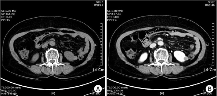
Fig. 1 A 62-year-old woman with a category II cystic lesion (hyperdense cyst) in the left kidney. The patient underwent laparoscopic partial nephrectomy. The final pathological diagnosis is renal cell carcinoma (papillary renal cell carcinoma). (A) Unenhanced computed tomography (CT) scan shows small, smooth-walled, high-density left renal cyst. (B) CT scan after administration of contrast material shows no enhancement of the cyst.
Reference
-
1. Bosniak MA. The current radiological approach to renal cysts. Radiology. 1986; 158:1–10.2. Smith AD, Remer EM, Cox KL, Lieber ML, Allen BC, Shah SN, et al. Bosniak category IIF and III cystic renal lesions: outcomes and associations. Radiology. 2012; 262:152–160.3. Remzi M, Marberger M. Renal tumor biopsies for evaluation of small renal tumors: why, in whom, and how? Eur Urol. 2009; 55:359–367.4. O'Malley RL, Godoy G, Hecht EM, Stifelman MD, Taneja SS. Bosniak category IIF designation and surgery for complex renal cysts. J Urol. 2009; 182:1091–1095.5. Israel GM, Bosniak MA. An update of the Bosniak renal cyst classification system. Urology. 2005; 66:484–488.6. Graumann O, Osther SS, Osther PJ. Characterization of complex renal cysts: a critical evaluation of the Bosniak classification. Scand J Urol Nephrol. 2011; 45:84–90.7. Curry NS, Cochran ST, Bissada NK. Cystic renal masses: accurate Bosniak classification requires adequate renal CT. AJR Am J Roentgenol. 2000; 175:339–342.8. Goenka AH, Remer EM, Smith AD, Obuchowski NA, Klink J, Campbell SC. Development of a clinical prediction model for assessment of malignancy risk in Bosniak III renal lesions. Urology. 2013; 82:630–635.9. Park HS, Jeong KS, Cheon J, Yoon DK, Jeong KB. The clinical significance of Bosniak classification in cystic renal masses : usefulness of preoperative computerized tomography in cystic renal masses. Korean J Urol. 1994; 35:498–503.10. Warren KS, McFarlane J. The Bosniak classification of renal cystic masses. BJU Int. 2005; 95:939–942.11. Benjaminov O, Atri M, O'Malley M, Lobo K, Tomlinson G. Enhancing component on CT to predict malignancy in cystic renal masses and interobserver agreement of different CT features. AJR Am J Roentgenol. 2006; 186:665–672.12. Silverman SG, Mortele KJ, Tuncali K, Jinzaki M, Cibas ES. Hyperattenuating renal masses: etiologies, pathogenesis, and imaging evaluation. Radiographics. 2007; 27:1131–1143.13. Vikram R, Ng CS, Tamboli P, Tannir NM, Jonasch E, Matin SF, et al. Papillary renal cell carcinoma: radiologic-pathologic correlation and spectrum of disease. Radiographics. 2009; 29:741–754.14. Silverman SG, Israel GM, Herts BR, Richie JP. Management of the incidental renal mass. Radiology. 2008; 249:16–31.15. Han HH, Choi KH, Oh YT, Yang SC, Han WK. Differential diagnosis of complex renal cysts based on lesion size along with the Bosniak renal cyst classification. Yonsei Med J. 2012; 53:729–733.16. Kouba E, Smith A, McRackan D, Wallen EM, Pruthi RS. Watchful waiting for solid renal masses: insight into the natural history and results of delayed intervention. J Urol. 2007; 177:466–470.17. Volpe A, Panzarella T, Rendon RA, Haider MA, Kondylis FI, Jewett MA. The natural history of incidentally detected small renal masses. Cancer. 2004; 100:738–745.18. Song C, Min GE, Song K, Kim JK, Hong B, Kim CS, et al. Differential diagnosis of complex cystic renal mass using multiphase computerized tomography. J Urol. 2009; 181:2446–2450.19. Weibl P, Klatte T, Waldert M, Remzi M. Complex renal cystic masses: current standards and controversies. Int Urol Nephrol. 2012; 44:13–18.
- Full Text Links
-
- Actions
-
Cited
- CITED
-
- Close
- Share
-
- Similar articles
-
- The Clinical Significance of Bosniak Classification in Cystic Renal Masses : Usefulness of Preoperative Computerized Tomography in Cystic Renal Masses
- Usefulness of the Bosniak Classification in Cystic Renal Mass on CT
- Collateral Circulation on the Neck after Common Carotid Ligation(Bosniak's Plexus)
- Differential Diagnosis of Complex Renal Cysts Based on Lesion Size along with the Bosniak Renal Cyst Classification
- Imaging Diagnosis and Management of Cystic Renal Masses: Introduction of an Update Proposal Bosniak Classification Version 2019
